. 2015 Jun 3;35(22):8493-506.
doi: 10.1523/JNEUROSCI.0377-15.2015.

The Chromatin Remodeling Protein Bptf Promotes Posterior Neuroectodermal Fate by Enhancing Smad2-Activated wnt8a Expression

Affiliations

The Chromatin Remodeling Protein Bptf Promotes Posterior Neuroectodermal Fate by Enhancing Smad2-Activated wnt8a Expression

Yuanqing Ma et al. J Neurosci. .

Abstract

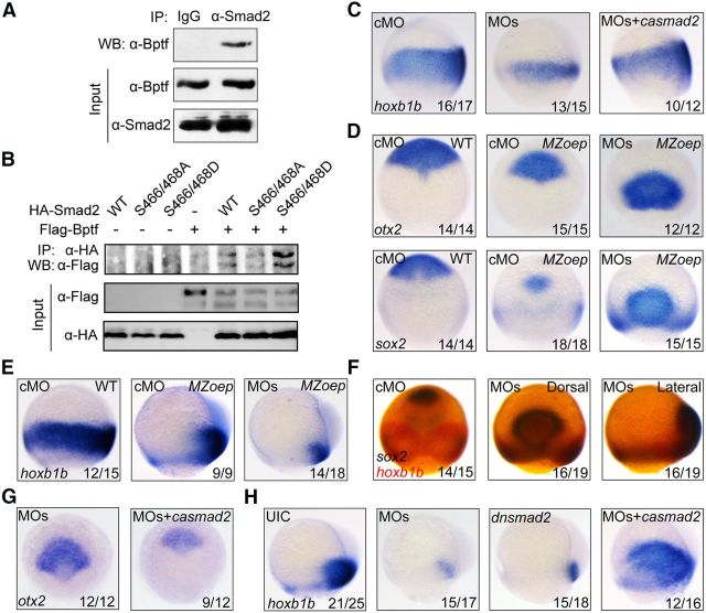
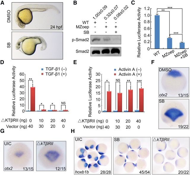
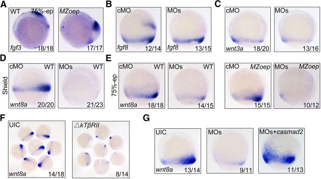
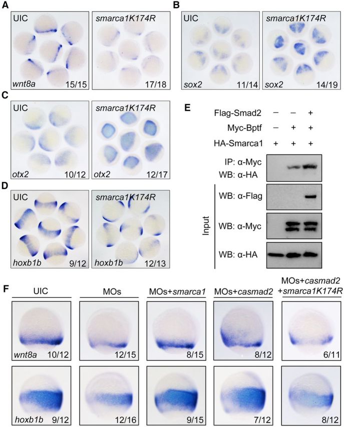
During vertebrate embryogenesis, the neuroectoderm is induced from dorsal ectoderm and then partitioned into anterior and posterior neuroectodermal domains by posteriorizing signals, such as Wnt and fibroblast growth factor. However, little is known about epigenetic regulation of posteriorizing gene expression. Here, we report a requirement of the chromatin remodeling protein Bptf for neuroectodermal posteriorization in zebrafish embryos. Knockdown of bptf leads to an expansion of the anterior neuroectoderm at the expense of the posterior ectoderm. Bptf functionally and physically interacts with p-Smad2, which is activated by non-Nodal TGF-β signaling, to promote the expression of wnt8a, a critical gene for neural posteriorization. Bptf and Smad2 directly bind to and activate the wnt8a promoter through recruiting NURF remodeling complex. When bptf function or TGF-β signal transduction is inhibited, the nucleosome density on the wnt8a promoter is increased. We propose that Bptf and TGF-β/Smad2 mediate nucleosome remodeling to regulate wnt8a expression and hence neural posteriorization.

Keywords: bptf; neural posteriorization; nucleosome remodeling; smad2; wnt8a.


Figures

Figure 1.
Figure 1.
Zebrafish bptf is expressed during early embryonic development and involved in head patterning. A, A′, Expression pattern of zebrafish bptf at indicated stages, detected by in situ hybridization with antisense (A, A′) or sense (A) probe. From one-cell stage to 32-cell stage, animal views; from sphere to bud stages, lateral views with dorsal to the right; 10-somite stage, dorsal view with anterior to the top; 24 and 30 hpf, lateral views with anterior to the left. B, Western blot analysis of endogenous Bptf protein expression during zebrafish embryo development. C, Effectiveness of bptf MO1. Embryos coinjected with 50 pg bptf-GFP plasmid DNA and 4 ng cMO or MO1 at one-cell stage. Green fluorescence was detected at 75% epiboly stage. MO1-injected embryos showed a noticeable decrease in green fluorescence compared with control embryos. D, Effectiveness of bptf MO2. The specific splicing-interfering MO (bptf MO2, targeting the splicing region between exon1 and intron1) interfered bptf mRNA product is only detected in morphants, whereas endogenous mRNA level was significantly deceased. β-actin is used as a control. E, Embryos were injected with 4 ng bptf MO1, 4 ng bptf MO2, or their mix (bptf MOs, each 4 ng) and harvested for Western blotting at 75% epiboly stage. UIC, Uninjected control. Band densities were analyzed using Quantity One software. The numbers on top of each lane indicate the relative band densities and the mean ± SD of Bptf after normalization to β-actin from 4 independent experiments. F, Morphological effects of bptf MO-injected wild-type or p53 mutant embryos at 24 hpf. Note the enlarged forebrain in bptf morphants (arrowhead). The ratio of embryos with the representative phenotypes was indicated. Scale bar, 200 μm.
Figure 2.
Figure 2.
bptf has crucial functions in neural posteriorization. A–E, Knockdown of bptf by morpholino injection blocks neural posteriorization. In situ hybridization of the anterior neural markers otx2 and sox2 (A, B, dorsal view with anterior at the top), the posterior neural marker hoxb1b, and the pan-neural marker sox3 (C, E, lateral views with dorsal to the right) at 75% epiboly stage. The expression of otx2 and sox2 in embryos injected with control MO or bptf MOs was analyzed at 75% epiboly stage by real-time PCR. *p < 0.05 (Student's t test). ***p < 0.001 (Student's t test). D, F–H, Knockdown of bptf by siRNA injection results in severe neural patterning defects. Embryos injected with 1.2 ng of indicated siRNAs at one-cell stage and harvested at 75% epiboly stage for real-time PCR (F) and in situ hybridization (G, H) analysis. NS, Nonsignificant. *p < 0.05 (Student's t test). F, I, The expression pattern of hoxb1b was analyzed by in situ hybridization in control MO and bptf MO-injected p53 mutant embryos at 75% epiboly stage. J, K, Abrogation of bptf function results in enlarged forebrain and loss of spinal cord. The expression pattern of six3 and shh was analyzed by in situ hybridization in control MO and bptf MO-injected embryos at the bud stage. J, Anterior view with dorsal at the top. K, Dorsal view, anterior is upward. L, M, Rescue of bptf morphants by hBptf. The expression of sox2 (L) and hoxb1b (M) at 75% epiboly stage in wild-type embryos injected with bptf MOs alone or together with 50 pg plasmid construct expressing hBptf. Black arrowheads indicate the representative spotted expression of hoxb1b.
Figure 3.
Figure 3.
Bptf associates with Smad2 to posteriorize the neuroectoderm independent of Nodal signaling. A, Zebrafish Bptf interacts with Smad2. Embryos were harvested and lysed at the 75% epiboly stage for coimmunoprecipitation using indicated antibodies. B, Bptf shows a much stronger binding affinity for activated Smad2. HEK293T cells were transfected with indicated expression plasmids encoding Flag-tagged Bptf, HA-tagged wild-type (WT) Smad2, phospho-resistant Smad2(S466/468A) mutant, and phospho-mimetic Smad2(S466/468D) mutant. Cells were harvested 48 h after transfection for immunoprecipitation with anti-HA antibody. C–E, Expression of neuroectodermal markers at 75% epiboly stage. Wild-type or MZoep embryos were injected with cMO or bptf MOs alone or together with casmad2 mRNA at the one-cell stage and harvested later for probing with indicated probes. Embryos were shown in lateral view (C,E) or dorsal view (D) with anterior to the top. F, Double in situ hybridization detection of sox2 (dark blue) and hoxb1b (red) in MZoep mutants injected with cMO or bptf MOs. The first two embryos were shown in dorsal view with anterior to the top and the last one in lateral view with dorsal to the right. G, H, The expression pattern of otx2 (G) and hoxb1b (H) in MZoep embryos. Injection doses: cMO, 8 ng; bptf MOs, 8 ng; casmad2 mRNA, 50 pg; dnsmad2 mRNA, 600 pg. Dorsal (G) or lateral views (H) are shown.
Figure 4.
Figure 4.
Inactivation of TGF-β signaling in MZoep mutant embryos leads to severe defects in neural AP patterning. A, Live embryos at 24 hpf. Embryos were treated with DMSO or 50 μm SB431542 (SB) from 16-cell stage to 24 hpf. The anterior is to the left. B, Detection of p-Smad2 by Western blotting in wild-type and MZoep mutant embryos. Embryos (WT and MZoep) were treated with DMSO or 50 μm SB431542 at 16-cell stage and harvested at 75% epiboly stage for Western blotting. Band densities were analyzed using Quantity One software. The numbers on top of each lane indicate the relative band densities and the mean ± SD of p-Smad2 after normalization to β-actin from 3 independent experiments. C, Reduction of ARE-luciferase reporter expression in MZoep mutant embryos treated with SB431542. WT or MZoep embryos were injected with the reporter plasmids at one-cell stage, treated with DMSO or SB431542 at 16-cell stage, and harvested at 75% epiboly stage for luciferase activity analysis. **p < 0.01 (Student's t test). ***p < 0.001 (Student's t test). D, E, ΔkTβRII overexpression specifically attenuates TGF-β-induced expression of ARE-luciferase. HEK293 cells were cotransfected with ARE-luciferase and FoxH1 constructs along with increasing amounts of ΔkTβRII construct, and treated with TGF-β1 (1 ng/ml) (D) or Activin A (10 ng/ml) (E) for 16 h before harvest for luciferase assay. *p < 0.05 (Student's t test), **p < 0.01 (Student's t test), ***p < 0.001 (Student's t test); NS, nonsignificant. F–H, Change of expression pattern of otx2 and hoxb1b at the 75% epiboly stage in MZoep embryos treated with 50 μm SB431542 or injected with 400 pg ΔkTβRII mNRA. Single embryos were shown in dorsal view for otx2 expression (F, G) and a group of embryos were shown in dorsal view mostly with posterior to the middle for hoxb1b expression (H).
Figure 5.
Figure 5.
Identification of Bptf and TGF-β/Smad2 coregulated genes during neural posteriorization. A–C, The expression pattern of fgf3, fgf8a, and wnt3a at 75% epiboly stages in wild-type, MZoep mutant, and bptf MOs injected embryos. All embryos were shown in lateral view with dorsal to the right. D, E, The expression of wnt8a is regulated by bptf, but not Nodal, signaling. wnt8a expression was assessed at shield (D) and 75% epiboly stages (E) by in situ hybridization in wild-type (D, E) and MZoep (E) embryos injected with cMO or bptf MOs. F, Reduction of wnt8a expression in ΔkTβRII mRNA-injected MZoep mutant embryos. A group of embryos at 75% epiboly stage were shown. G, Ectopic expression of casmad2 restores bptf MOs-induced decrease of wnt8a expression. MZoep mutant embryos were injected with 8 ng bptf MOs alone or together with 50 pg casmad2 mRNA at the one-cell stage and harvested at 75% epiboly stage for in situ hybridization.
Figure 6.
Figure 6.
Bptf and TGF-β/Smad2 posteriorize the neuroectoderm via wnt8a. A, Wnt/β-catenin signaling is reduced in bptf morphants. gfp expression was examined at shield and 75% epiboly stages by in situ hybridization in TOPdGFP transgenic fish embryos injected with cMO or bptf MOs. Top, Animal-pole views with dorsal to the right. Bottom, Lateral views with dorsal to the right. B, Expression of otx2 and hoxb1b at 75% epiboly stage in wild-type embryos injected with bptf MOs alone or together with 25 pg wnt8a mRNA. Injection of wnt8a mRNA could compromise the expansion of the anterior neuroectoderm and the loss of posterior neuroectoderm in bptf morphants. C, D, Neural patterning defects resulting from inactivation of both bptf and TGF-β signaling were rescued by coinjection of wnt8a mRNA. Wild-type embryos (C) or MZoep mutants (D) were injected with indicated MOs or RNAs at the one-cell stage and harvested at 75% epiboly stage for in situ hybridization using otx2 and hoxb1b probes. Injection doses were as follows: bptf MOs, 8 ng; ▵kTβRII mRNA, 400 pg; wnt8a mRNA, 25 pg.
Figure 7.
Figure 7.
Bptf and Smad2 regulate wnt8a expression through distinct cis-elements. A, Schematic illustration of wnt8a promoter-driven luciferase reporters. B, C, The Bptf-binding element is essential for wnt8a transcription. Wild-type embryos were injected with indicated MOs and wnt8a promoter-driven reporter constructs at one-cell stage and then harvested and lysed at 75% epiboly stage for luciferase activity analysis. The −1101 to −924 promoter region is important for wnt8a transcription (B). 1101-mb-P-luc showed a much lower luciferase activity than the corresponding wild-type reporter (C). D, The −924 to −657 region of the wnt8a promoter is essential for responding to casmad2 overexpression. The indicated constructs and casmad2 mRNA were injected into embryos at one-cell stage, and the relative luciferase activity was measured at 75% epiboly stage. E, The Smad2/4-binding site is essential for responding to casmad2 overexpression. 1101-ms-P-luc or the related wild-type reporter 1101-P-luc was injected into embryos, and their responsiveness to overexpressed casmad2 was measured. Data represent the mean ± SD for triplicate experiments. *p < 0.05 (Student's t test). **p < 0.01 (Student's t test). ***p < 0.001 (Student's t test). F, Bptf and Smad2 specifically bind to the wnt8a promoter. ChIP assays were performed with control IgG and antibodies against Bptf or Smad2/3 in wild-type embryos. The immunoprecipitated DNA was amplified by semiqPCR with the primers recognizing specific regions denoted in the top. wnt8a up, a region upstream of the wnt8a TSS, which contains Bptf and Smad2/4 binding motifs; wnt8a down, a region downstream of the wnt8a TSS. G, ChIP experiments were performed in wild-type, ΔkTβRII mRNA-injected, or SB431542-treated embryos using anti-Bptf antibody. The immunoprecipitated DNA was amplified by qPCR with primers to detect the region upstream of the wnt8a TSS. NS, Nonsignificant. H, I, Wild-type embryos were injected with cMO or bptf MOs at the one-cell stage and harvested at 75% epiboly stage for ChIP experiments with anti-Bptf (H) or anti-Smad2/3 (I) antibodies. The immunoprecipitated DNA was subjected to qPCR with primers indicated in G.
Figure 8.
Figure 8.
Smarca1 is required for wnt8a expression and neural posteriorization. A, Interference in smarca1 inhibits wnt8a expression. Uninjected and smarca1K174R mRNA-injected (125 pg) embryos were probed at 75% epiboly stage for wnt8a expression by in situ hybridization. B–D, smarca1K174R overexpression disrupts neuroectodermal AP patterning. One-cell stage embryos were injected with 125 pg smarca1K174R mRNA and harvested at 75% epiboly stage for in situ hybridization to detect the anterior neural markers sox2 (B) and otx2 (C) and the posterior neural marker hoxb1b (D). E, Smad2 enhances the association of Bptf and Smarca1. HEK293T cells were transfected with combinations of plasmids expressing Myc-Bptf, HA-Smarca1, and Flag-Smad2 as indicated. After 48 h, cells were lysed for immunoprecipitation with anti-Myc antibody. F, Smarca1 functions on Bptf- and Smad2-mediated wnt8a expression and neural posteriorization. Embryos were injected with indicated MOs and mRNAs at the one-cell stage and harvested at 75% epiboly stage for in situ hybridization to detect alteration of wnt8a and hoxb1b expression. Embryos were orientated laterally with dorsal to the right. Injection doses are as follows: bptf MOs, 8 ng; smarca1 mRNA, 250 pg; smarca1K174R mRNA, 125 pg; casmad2 mRNA, 50 pg.
Figure 9.
Figure 9.
Inactivation of bptf or TGF-β signaling induces nucleosome repositioning within the wnt8a promoter. A, MNase digestion of chromatin isolated from embryos at 75% epiboly stage. Digestion with 320 units per milliliters of MNase for 30 min was appropriate to produce mononucleosome-sized DNAs. B, C, The dynamic changes of nucleosomal positions at the wnt8a promoter in bptf morphants (B) or ΔkTβRII-overexpressing embryos (C). There were five positioned nucleosomes (N1, N2, N3, N4, and N5) within the −1449 to −416 region of the wnt8a promoter in cMO-injected embryos. Bptf (green) and Smad2 (red) binding motifs were located in the DNA sequences occupied by N3. A solid increase in DNA amount was detected at N3 positioning site in bptf morphants and ΔkTβRII-overexpressing embryos. NS, Nonsignificant. **p < 0.01 (Student's t test). ***p < 0.001 (Student's t test). B′, C′, Injection of bptf MOs or ΔkTβRII mRNA results in an increase in histone density over the positioned N3 within the wnt8a promoter. Embryos were injected with 8 ng bptf MOs or 400 pg ΔkTβRII mRNA at one-cell stage and harvested at 75% epiboly stage. The resultant chromatin was extracted and subjected to ChIP assays with anti-H3 antibody followed by qPCR. NS, Nonsignificant. *p < 0.05 (Student's t test).

References

    1. Appel B. Zebrafish neural induction and patterning. Dev Dyn. 2000;219:155–168. doi: 10.1002/1097-0177(2000). - DOI - PubMed
    1. Badenhorst P, Xiao H, Cherbas L, Kwon SY, Voas M, Rebay I, Cherbas P, Wu C. The Drosophila nucleosome remodeling factor NURF is required for Ecdysteroid signaling and metamorphosis. Genes Dev. 2005;19:2540–2545. doi: 10.1101/gad.1342605. - DOI - PMC - PubMed
    1. Barak O, Lazzaro MA, Lane WS, Speicher DW, Picketts DJ, Shiekhattar R. Isolation of human NURF: a regulator of Engrailed gene expression. EMBO J. 2003;22:6089–6100. doi: 10.1093/emboj/cdg582. - DOI - PMC - PubMed
    1. Brand T, MacLellan WR, Schneider MD. A dominant-negative receptor for type beta transforming growth factors created by deletion of the kinase domain. J Biol Chem. 1993;268:11500–11503. - PubMed
    1. Buckles GR, Thorpe CJ, Ramel MC, Lekven AC. Combinatorial Wnt control of zebrafish midbrain-hindbrain boundary formation. Mech Dev. 2004;121:437–447. doi: 10.1016/j.mod.2004.03.026. - DOI - PubMed

Publication types

MeSH terms