Multicenter Study
. 2015 Jun 3;35(22):8672-82.
doi: 10.1523/JNEUROSCI.0862-15.2015.

Coupled changes in brain white matter microstructure and fluid intelligence in later life

Affiliations
Multicenter Study

Coupled changes in brain white matter microstructure and fluid intelligence in later life

Stuart J Ritchie et al. J Neurosci. .

Abstract

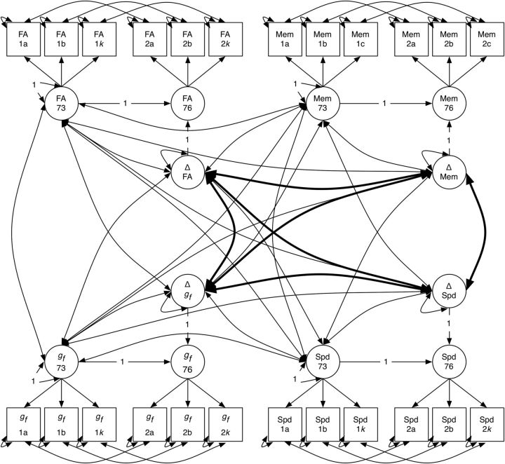
Understanding aging-related cognitive decline is of growing importance in aging societies, but relatively little is known about its neural substrates. Measures of white matter microstructure are known to correlate cross-sectionally with cognitive ability measures, but only a few small studies have tested for longitudinal relations among these variables. We tested whether there were coupled changes in brain white matter microstructure indexed by fractional anisotropy (FA) and three broad cognitive domains (fluid intelligence, processing speed, and memory) in a large cohort of human participants with longitudinal diffusion tensor MRI and detailed cognitive data taken at ages 73 years (n = 731) and 76 years (n = 488). Longitudinal changes in white matter microstructure were coupled with changes in fluid intelligence, but not with processing speed or memory. Individuals with higher baseline white matter FA showed less subsequent decline in processing speed. Our results provide evidence for a longitudinal link between changes in white matter microstructure and aging-related cognitive decline during the eighth decade of life. They are consistent with theoretical perspectives positing that a corticocortical "disconnection" partly explains cognitive aging.

Keywords: cognitive aging; diffusion tensor imaging; fluid intelligence; fractional anisotropy; processing speed; white matter microstructure.


Figures

Figure 1.
Figure 1.
Examples of major white matter tracts segmented using probabilistic neighborhood tractography overlaid on fractional anisotropy maps for a representative subject; segmented white matter tracts are shown in orange, with seed points indicated with a green cross. Top row (left to right), Genu of corpus callosum, splenium of corpus callosum, and anterior thalamic radiation. Middle row (left to right), rostral cingulum and inferior longitudinal fasciculus. Bottom row (left to right), Arcuate fasciculus and uncinate fasciculus. The latter five tracts were measured for both the left and right hemispheres, giving a total of 12 tracts. Tracts were measured and processed in an identical fashion at both the age ∼73 years and age ∼76 years measurement waves.
Figure 2.
Figure 2.
Full path diagram for the four-way multivariate latent difference score model. For each variable, two latent factors (circles; age 73 and 76 years) are estimated from the manifest indicators (squares, 1a-k and 2a-k), and a latent change factor is calculated from the change between age 73 and 76 years (circles, Δ). Bolded paths indicate change–change correlations, the main paths of interest in the present study. For space reasons, only three manifest variables are shown for each latent factor; in the model: FA, gf, and processing speed were all estimated using more than three indicators (12, 4, and 4, respectively). Paths labeled “1” were fixed to 1 to identify the model and estimate the latent change factor. For clarity, the model mean structure is not shown. Mem, Memory; Spd, processing speed.
Figure 3.
Figure 3.
Results from key parts of the latent difference score model. The full model is shown in Figure 2. Full results with significance levels are shown in Table 3 (measurement models; i.e., loadings of each test/tract on its general factor) and Table 4 (structural model; i.e., associations among the baseline and change latent traits). Correlations among latent variables (circles) of brain-wide white matter FA, gf, processing speed (Spd), and memory (Mem) are shown as values on each path (line), with SE in parentheses. Dashed paths are not statistically significant. a, All four latent variables were correlated significantly and positively at baseline. b, Baseline FA was correlated with the change in processing speed between ages ∼73 and ∼76 years, but not with the change in gf or memory. c, Change (Δ) in FA and gf were positively correlated. Changes in gf, Spd, and Mem were all significantly and strongly correlated.

References

    1. Aribisala BS, Valdés Hernández MC, Royle NA, Morris Z, Muñoz Maniega S, Bastin ME, Deary IJ, Wardlaw JM. Brain atrophy associations with white matter lesions in the ageing brain: the Lothian Birth Cohort 1936. Eur Radiol. 2013;23:1084–1092. doi: 10.1007/s00330-012-2677-x. - DOI - PubMed
    1. Bastin ME, Muñoz Maniega S, Ferguson KJ, Brown LJ, Wardlaw JM, MacLullich AM, Clayden JD. Quantifying the effects of normal ageing on white matter structure using unsupervised tract shape modelling. Neuroimage. 2010;51:1–10. doi: 10.1016/j.neuroimage.2010.02.036. - DOI - PMC - PubMed
    1. Behrens TE, Berg HJ, Jbabdi S, Rushworth MF, Woolrich WM. Probabilistic diffusion tractography with multiple fibre orientations: what can we gain? Neuroimage. 2007;34:144–155. doi: 10.1016/j.neuroimage.2006.09.018. - DOI - PMC - PubMed
    1. Bennett IJ, Madden DJ. Disconnected aging: cerebral white matter integrity and age-related differences in cognition. Neuroscience. 2014;276:187–205. doi: 10.1016/j.neuroscience.2013.11.026. - DOI - PMC - PubMed
    1. Booth T, Bastin ME, Penke L, Maniega SM, Murray C, Royle NA, Gow AJ, Corley J, Henderson RD, Starr JM, Wardlaw JM, Deary IJ. Brain white matter tract integrity and cognitive abilities in community-dwelling older people: the Lothian Birth Cohort, 1936. Neuropsychology. 2013;27:595–607. doi: 10.1037/a0033354. - DOI - PMC - PubMed

Publication types

LinkOut - more resources