. 2015 Aug 1;26(15):2823-32.
doi: 10.1091/mbc.E15-01-0009. Epub 2015 Jun 3.

Myristoylated CIL-7 regulates ciliary extracellular vesicle biogenesis

Affiliations

Myristoylated CIL-7 regulates ciliary extracellular vesicle biogenesis

Julie E Maguire et al. Mol Biol Cell. .

Abstract

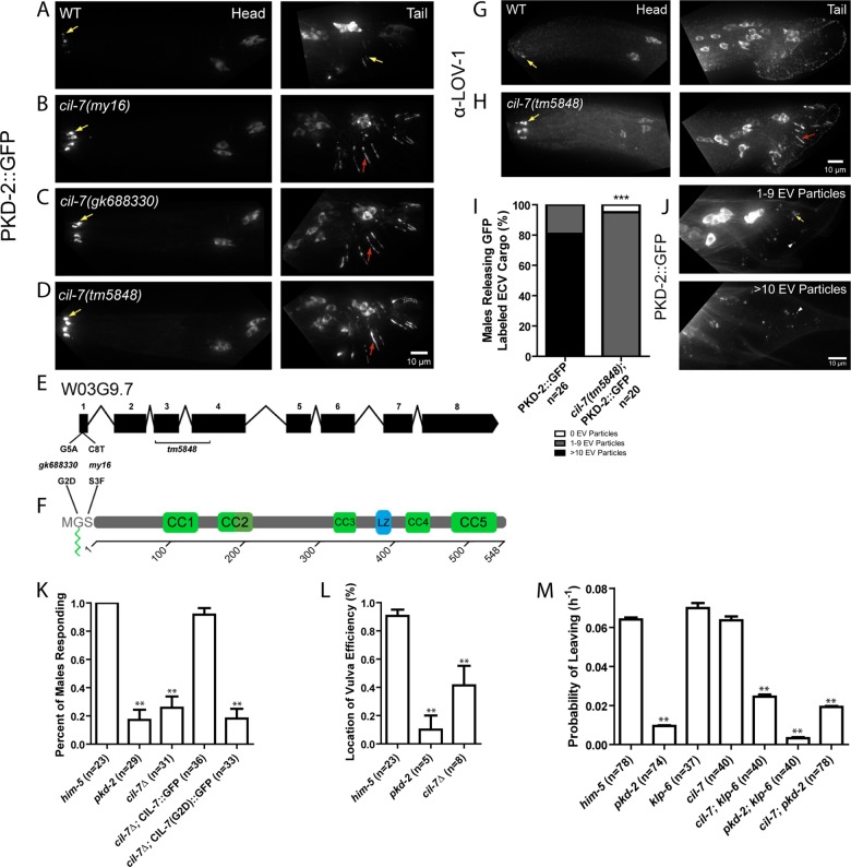
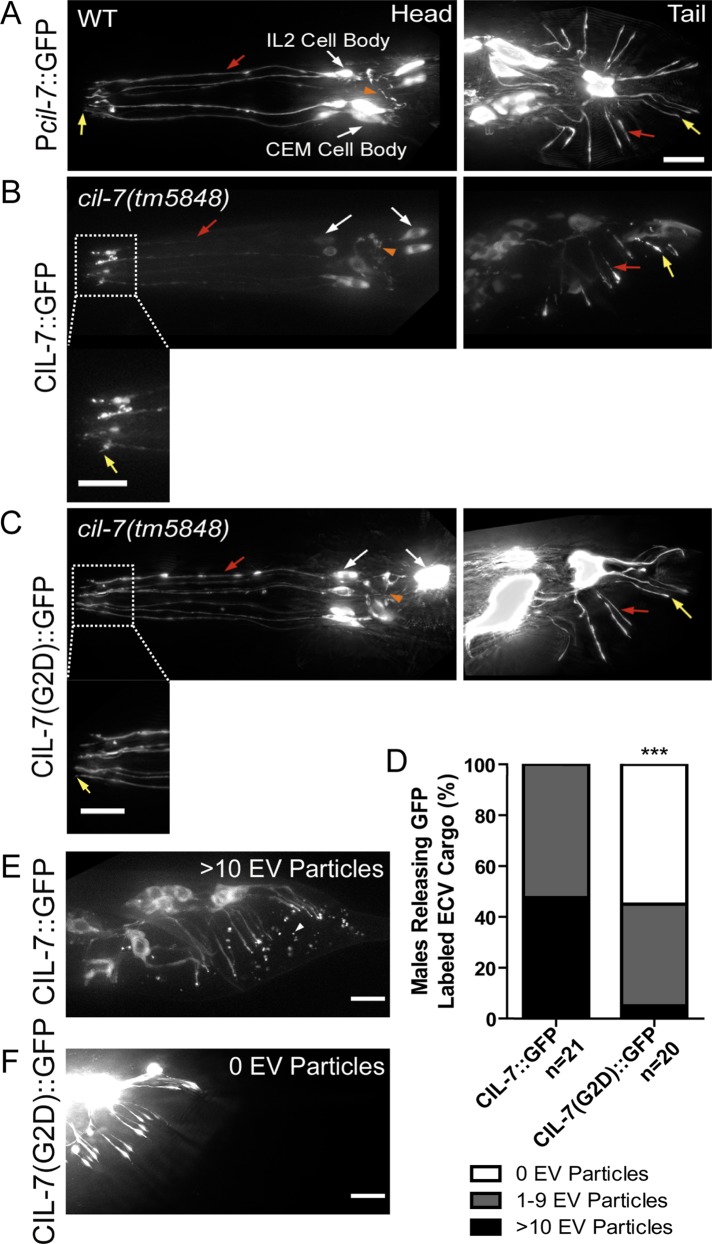
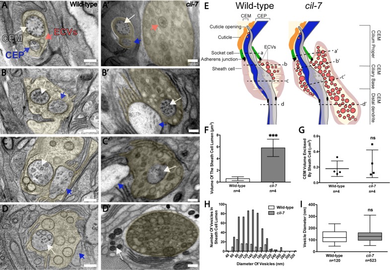
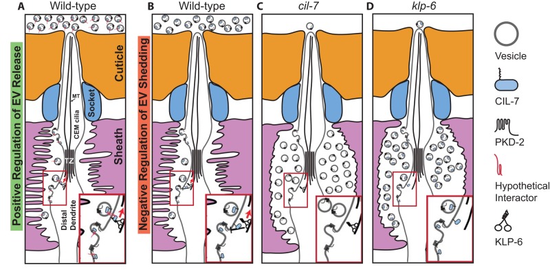
The cilium both releases and binds to extracellular vesicles (EVs). EVs may be used by cells as a form of intercellular communication and mediate a broad range of physiological and pathological processes. The mammalian polycystins (PCs) localize to cilia, as well as to urinary EVs released from renal epithelial cells. PC ciliary trafficking defects may be an underlying cause of autosomal dominant polycystic kidney disease (PKD), and ciliary-EV interactions have been proposed to play a central role in the biology of PKD. In Caenorhabditis elegans and mammals, PC1 and PC2 act in the same genetic pathway, act in a sensory capacity, localize to cilia, and are contained in secreted EVs, suggesting ancient conservation. However, the relationship between cilia and EVs and the mechanisms generating PC-containing EVs remain an enigma. In a forward genetic screen for regulators of C. elegans PKD-2 ciliary localization, we identified CIL-7, a myristoylated protein that regulates EV biogenesis. Loss of CIL-7 results in male mating behavioral defects, excessive accumulation of EVs in the lumen of the cephalic sensory organ, and failure to release PKD-2::GFP-containing EVs to the environment. Fatty acylation, such as myristoylation and palmitoylation, targets proteins to cilia and flagella. The CIL-7 myristoylation motif is essential for CIL-7 function and for targeting CIL-7 to EVs. C. elegans is a powerful model with which to study ciliary EV biogenesis in vivo and identify cis-targeting motifs such as myristoylation that are necessary for EV-cargo association and function.


Figures

FIGURE 1:
FIGURE 1:
cil-7 is required for the localization of the PCs and for male mating behaviors. (A) PKD-2::GFP localized to the cilia and cell bodies of the CEM, RnB, and HOB neurons of males. (B–D) In cil-7(my16), cil-7(gk688330) and cil-7(tm5848) males, head CEM neurons and tail HOB and RnB neurons accumulated PKD-2::GFP along the dendrites and cilia. (E) cil-7 genomic structure. my16 (C8 → T) and gk688330 (G5 → A) are missense SNPs. tm5848 is an out-of-frame deletion. (F) cil-7 encodes a predicted protein with an N-terminal myristoylation motif, five coiled-coil domains, and a leucine zipper. (G) The anti–LOV-1 antibody showed that endogenous LOV-1 localized to the cilia and cell bodies of the CEM, HOB, and RnB neurons. (H) In cil-7(tm5848) males, endogenous LOV-1 accumulated at the base and along the cilia of the CEM neurons. Excess LOV-1 appeared along the cilia and the dendrites of the RnB neurons. (I) In the cuticle of L4 molting wild-type males, >10 PKD-2::GFP–containing EV particles were observed in 80% of animals scored. In cil-7(tm5848) L4 molting males, much fewer PKD-2::GFP–containing EVs were observed. (J) Representative images of PKD-2::GFP EV particle ranges (1–9 or >10) observed to be trapped in the molted tail cuticle of late L4 males. (K) cil-7(tm5848) males did not respond to hermaphrodite contact. The translational reporter Pcil-7::CIL-7::GFP rescued the response defect of cil-7(tm5848) males. By contrast, the myristoylation-defective reporter Pcil-7::CIL-7(G2D)::GFP failed to rescue response defects of cil-7(tm5848) males. (L) cil-7(tm5848) males were location-of-vulva (Lov) defective. (M) cil-7(tm5848) males were not-leaving-assay defective (nonLas). The cil-7(tm5848); klp-6(my8) double mutant was Las, unlike the cil-7(tm5848) or klp-6(my8) single mutants. In I, ***p < 0.0001 by the Mann–Whitney test. In K, data were analyzed with Fisher's exact test between all groups, followed by Holm–Bonferroni multiple comparison adjustment with a α of 0.01 (**). In L, data were analyzed with the pairwise Mann–Whitney U-test between all groups, followed by Holm–Bonferroni multiple comparison adjustment with a total α of 0.01 (**). In M, PL values were compared by Holm–Bonferroni multiple comparison adjustment with a total α of 0.01 (**). Yellow arrow, cilia; red arrow, dendrite; white arrowhead, EV particle.
FIGURE 2:
FIGURE 2:
cil-7 is expressed in the 27 EV-releasing ciliated sensory neurons and is targeted to EVs in a myristoylation motif–dependent manner. (A) Pcil-7::GFP was expressed in CEM, RnB, HOB, and IL2 neurons. (B) Pcil-7::CIL-7::GFP in cil-7(tm5848) males localized throughout neurons, including cilia, but was excluded from the nucleus. Pcil-7::CIL-7::GFP rescues cil-7(tm5848) defects (Figure 1K), indicating that this reporter is functional. (C) Pcil-7::CIL-7(G2D)::GFP in cil-7(tm5848) males localized throughout neurons, including cilia, but was excluded from the nucleus. Pcil-7::CIL-7(G2D)::GFP does not rescue cil-7(tm5848) defects (Figure 1K), indicating that this reporter is not functional. (D) CIL-7::GFP as EV cargo was trapped in the molted tail cuticle of the late L4 male in a range from either 1–9 EV particles or >10 EV particles per male tail. The CIL-7(G2D)::GFP myristoylation mutant is not efficiently targeted to EVs. (E) In 100% of animals, CIL-7::GFP was observed in the molted tail cuticle in varying ranges; depicted here is an example of >10 EV particles. (F) In <50% of animals, CIL-7 (G2D)::GFP was observed as EV cargo. In E, ***p < 0.0001 by the Mann–Whitney test. Yellow arrow, cilia; red arrow, dendrite; white arrow, cell body; orange arrowhead, axon; white arrowhead, EV particle.
FIGURE 3:
FIGURE 3:
cil-7 mutants accumulate EVs in the cephalic lumen but are not defective in ciliogenesis. (A–D) Series of TEM images in a wild-type animal displaying EVs in the sheath cell lumen that surrounds the CEM and CEP distal dendritic region (lumen is pseudocolored yellow). (A′–D′) TEM images revealed that cil-7(tm5848) males had excessive amounts of EVs in the sheath cell lumen. Both CEM and CEP axonemes appeared normal. (E) Cartoon depiction of the cil-7(tm5848) mutant phenotype compared with wild type, including increased quantity of EVs and distended sheath cell lumen. Dashed lines indicate the location of the A–D and A′–D′ TEM images with respect to the cephalic sensillum. (F) Compared to wild type (0.6 ± 0.3 μm3), the cil-7(tm5848) sheath cell lumen is 10 times larger (5.8 ± 1.5 μm3 [average volume ± SD]; n = number of cephalic sensilla). (G) The average CEM volume enclosed within the sheath cell that surrounds the CEM distal dendrite and axoneme in wild type was 0.2 ± 0.1 μm3, and the corresponding volume in cil-7(tm5848) was 0.24 ± 0.2 μm3 (average volume ± SD; n = number of cephalic sensilla), indicating that the integrity of CEM neurons is normal in cil-7 mutants. (H) Compared to wild type, cil-7(tm5848) animals displayed an increased number of EVs in the sheath cell lumen. The frequency distribution of EV sizes in cil-7(tm5848) was slightly favored toward EVs above the average mean (skewness of the diameter distribution as measured in Excel: wild type, 0.46; cil-7, 0.57). (I) The average EV diameter of wild-type and cil-7(tm5848) males was not significantly different. The average EV diameter for wild type was 129.1 ± 49.4 nm and the average vesicle diameter for cil-7(tm5848) was 131.8 ± 43.3 nm (average diameter ± SD). In F, ***p < 0.001 by the unpaired t test. In G, a nonsignificant difference was determined by the unpaired t test. In I, a nonsignificant difference was determined by Welch's t Test. Box-and-whisker plots are included in I, where the plot is divided into limits that are each 25% of the population.
FIGURE 4:
FIGURE 4:
Models for CIL-7– and KLP-6–mediated EV biogenesis. (A) KLP-6 and CIL-7 may be positive regulators of EV environmental release. In this model, EVs bud from the ciliary base and are shed into the cephalic lumen. From here, KLP-6 and IFT may propel EVs along the ciliary membrane surface to the cuticular opening. Myristoylated, membrane-associated CIL-7 is EV cargo that acts with an unidentified transmembrane tether. The tethered EV binds a ciliary surface protein that is transported by KLP-6 and IFT. Inset, the EV tethered to an unidentified protein such as a flagellar membrane glycoprotein. (B) CIL-7 and KLP-6 may act as dual negative regulators of EV shedding. Inset, a negative feedback loop with CIL-7 and KLP-6 acting in concert to regulate EV shedding. (C, D) In the absence of cil-7 or klp-6 as a positive regulator of EV release or negative regulator of EV shedding, the net result is an accumulation of EVs within the cephalic lumen. In both models, klp-6 acts within the EV-releasing ciliated sensory neurons, whereas cil-7 may act in the EV-releasing cell or in the EV itself. Insets of C and D depict EV shedding that is not regulated in the absence of CIL-7 and KLP-6, respectively. TZ, transition zone.

References

    1. Bae YK, Barr MM. Sensory roles of neuronal cilia: cilia development, morphogenesis, and function in C. elegans. Front Biosci. 2008;13:5959–5974. - PMC - PubMed
    1. Bae YK, Kim E, L'Hernault SW, Barr MM. The CIL-1 PI 5-phosphatase localizes TRP Polycystins to cilia and activates sperm in C. elegans. Curr Biol. 2009;19:1599–1607. - PMC - PubMed
    1. Bae YK, Lyman-Gingerich J, Barr MM, Knobel KM. Identification of genes involved in the ciliary trafficking of C. elegans PKD-2. Dev Dyn. 2008;237:2021–2029. - PMC - PubMed
    1. Bae YK, Qin H, Knobel KM, Hu J, Rosenbaum JL, Barr MM. General and cell-type specific mechanisms target TRPP2/PKD-2 to cilia. Development. 2006;133:3859–3870. - PubMed
    1. Bakeberg JL, Tammachote R, Woollard JR, Hogan MC, Tuan HF, Li M, van Deursen JM, Wu Y, Huang BQ, Torres VE, et al. Epitope-tagged Pkhd1 tracks the processing, secretion, and localization of fibrocystin. J Am Soc Nephrol. 2011;22:2266–2277. - PMC - PubMed

Publication types