

Automatic Identification of Cochlear Implant Electrode Arrays for Post-Operative Assessment
- PMID: 26041945
- PMCID: PMC4450802
- DOI: 10.1117/12.878490
Automatic Identification of Cochlear Implant Electrode Arrays for Post-Operative Assessment
Abstract
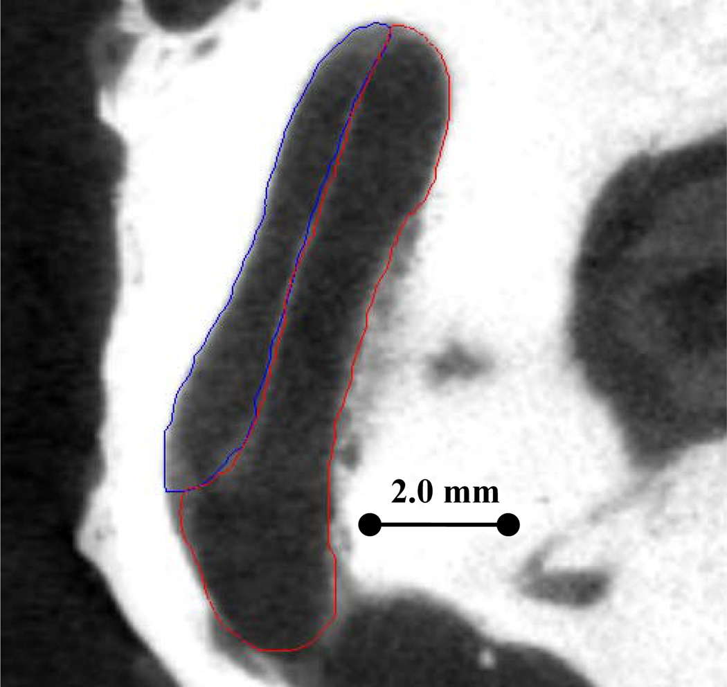
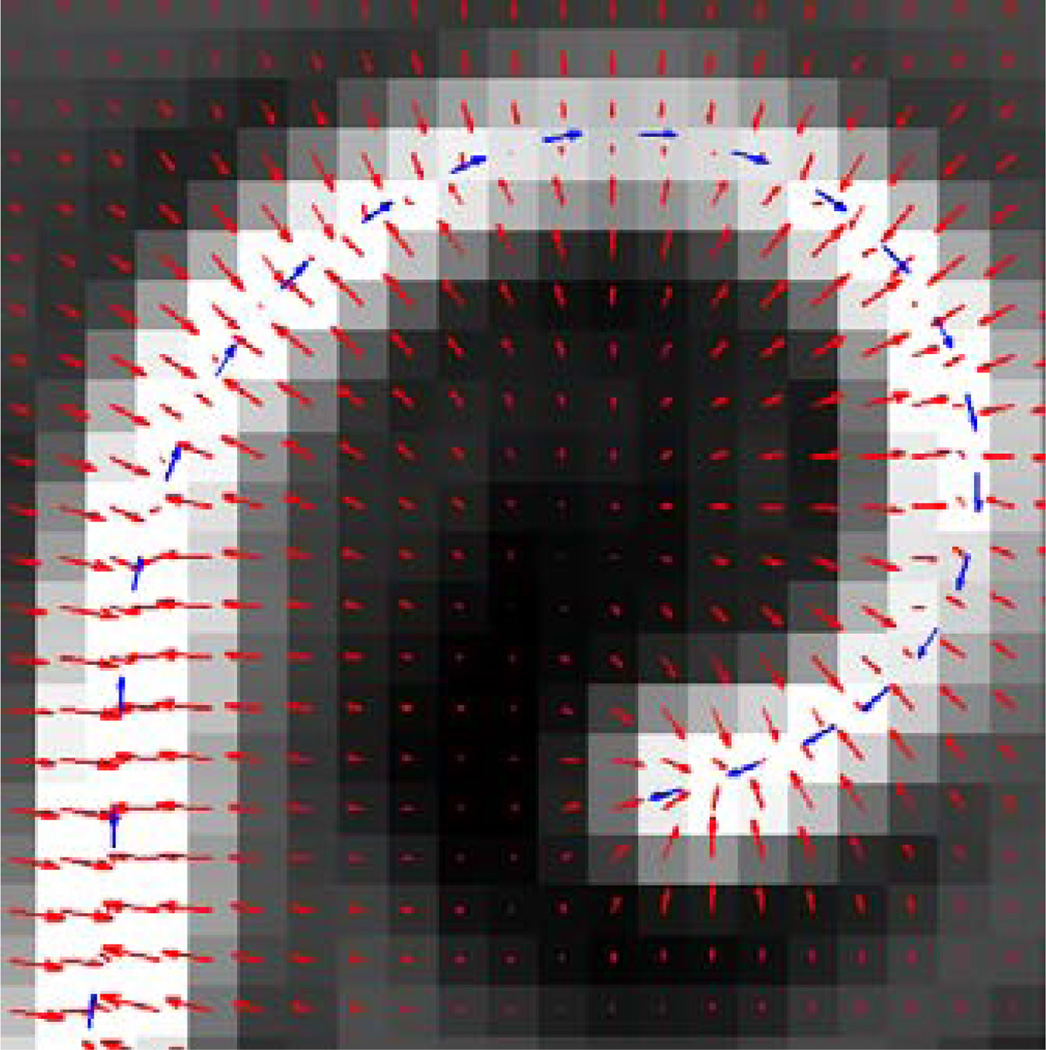
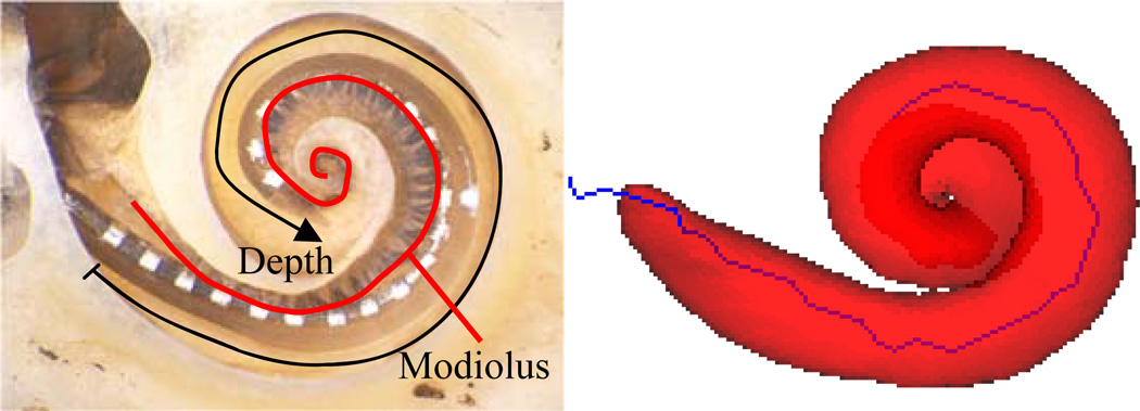
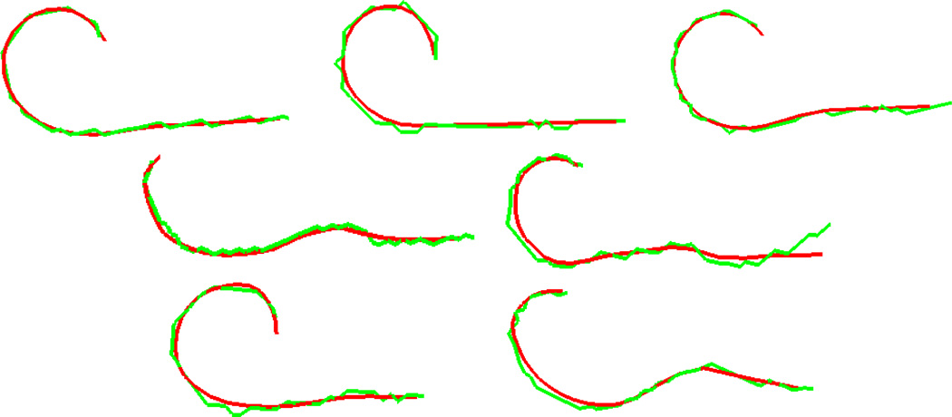
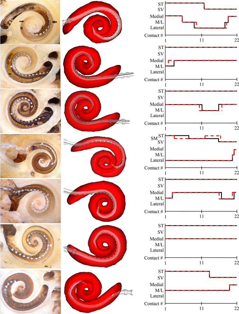
Cochlear implantation is a procedure performed to treat profound hearing loss. Accurately determining the postoperative position of the implant in vivo would permit studying the correlations between implant position and hearing restoration. To solve this problem, we present an approach based on parametric Gradient Vector Flow snakes to segment the electrode array in post-operative CT. By combining this with existing methods for localizing intra-cochlear anatomy, we have developed a system that permits accurate assessment of the implant position in vivo. The system is validated using a set of seven temporal bone specimens. The algorithms were run on pre- and post-operative CTs of the specimens, and the results were compared to histological images. It was found that the position of the arrays observed in the histological images is in excellent agreement with the position of their automatically generated 3D reconstructions in the CT scans.
Keywords: Cochlear Implant; Contour Advance; Snake Segmentation.
Figures
References
-
- Skinner MW, Holden TA, Whiting BR, et al. In vivo estimates of the position of advanced bionics electrode arrays in the human cochlea. Ann Otol Rhinol Laryngol Suppl. 2007 Apr;197:2–24. - PubMed
-
- Aschendorff A, Kromeier J, Klenzner T, Laszig R. Quality Control After Insertion of the Nucleus Contour and Contour Advance Electrode in Adults. Ear & Hearing. 2007 Apr;28:75S–79S. - PubMed
-
- Noble JH, Rutherford RB, Labadie RF, Majdani O, Dawant BM. Modeling and segmentation of intra-cochlear anatomy in conventional CT. Proceedings of the SPIE – Medical Imaging 2010: Image Processing. :762302.
-
- Xu C, Prince JL. Snakes, shapes, and gradient vector flow. IEEE Trans. on Image Processing. 1998;7(3):259–269. - PubMed
-
- Kass M, Witkin A, Terzopoulos D. Snakes: Active Contour Models. Int’l Jour. Of Computer Vision. 1988:321–331.
Grants and funding
LinkOut - more resources
Full Text Sources
Other Literature Sources
Research Materials