

Pathogenesis of hepatic encephalopathy and brain edema in acute liver failure
- PMID: 26041966
- PMCID: PMC4442857
- DOI: 10.1016/j.jceh.2014.02.004
Pathogenesis of hepatic encephalopathy and brain edema in acute liver failure
Abstract
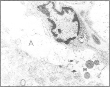
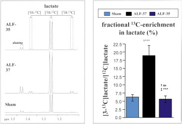
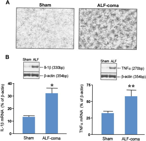
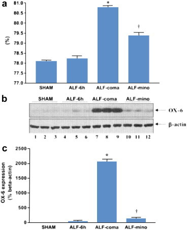
Neuropathologic investigations in acute liver failure (ALF) reveal significant alterations to neuroglia consisting of swelling of astrocytes leading to cytotoxic brain edema and intracranial hypertension as well as activation of microglia indicative of a central neuroinflammatory response. Increased arterial ammonia concentrations in patients with ALF are predictors of patients at risk for the development of brain herniation. Molecular and spectroscopic techniques in ALF reveal alterations in expression of an array of genes coding for neuroglial proteins involved in cell volume regulation and mitochondrial function as well as in the transport of neurotransmitter amino acids and in the synthesis of pro-inflammatory cytokines. Liver-brain pro-inflammatory signaling mechanisms involving transduction of systemically-derived cytokines, ammonia neurotoxicity and exposure to increased brain lactate have been proposed. Mild hypothermia and N-Acetyl cysteine have both hepato-protective and neuro-protective properties in ALF. Potentially effective anti-inflammatory agents aimed at control of encephalopathy and brain edema in ALF include etanercept and the antibiotic minocycline, a potent inhibitor of microglial activation. Translation of these potentially-interesting findings to the clinic is anxiously awaited.
Keywords: ALF, acute liver failure; ATP, adenosine triphosphate; BBB, blood-brain barrier; CCL2, chemokine ligand-2; CMRO2, cerebral metabolic rate for oxygen; CNS, central nervous system; EEG, electroencephalography; GABA, gamma-aminobutyric acid; GFAP, glial fibrillary acidic protein; IgG, immunoglobulin; MRS, magnetic resonance spectroscopy; NAC, N-Acetyl cysteine; NMDA, N-methyl-d-aspartate; SIRS, systemic inflammatory response syndrome; SNATs, several neutral amino acid transport systems; TLP, translocator protein; TNFα, tumor necrosis factor alpha; acute liver failure; hepatic encephalopathy; intracranial hypertension; microglial activation; neuroinflammation.
Figures
References
-
- Aggarwal S., Kramer D., Yonas H. Cerebral hemodynamic and metabolic changes in fulminant hepatic failure: a retrospective study. Hepatology. 1994;19:80–87. - PubMed
-
- Kato M., Hughes R.D., Keays R.T., Williams R. Electron microscopic study of brain capillaries in cerebral edema from fulminant hepatic failure. Hepatology. 1992;15:1060–1066. - PubMed
-
- Thumburu K.K., Dhiman R.K., Vasishta R.K. Expression of astrocytic genes coding for proteins implicated in neural excitation and brain edema is altered after acute liver failure. J Neurochem. 2014;128:617–627. - PubMed
-
- Potvin M., Morrison H.F., Hinchey E.J. Cerebral abnormalities in hepatectomised rats with acute hepatic coma. Lab Invest. 1984;50:560–564. - PubMed
-
- Chastre A., Belanger M., Nguyen B.N., Butterworth R.F. Lipo-polysaccharide precipitates hepatic encephalopathy and increases blood-brain barrier permeability in mice with acute liver failure. Liver Int. 2014;34:353–361. - PubMed
Publication types
LinkOut - more resources
Full Text Sources
Other Literature Sources
Miscellaneous
