Review
. 2015 May 18:8:14.
doi: 10.3389/fnmol.2015.00014. eCollection 2015.

Lentiviral vectors as tools to understand central nervous system biology in mammalian model organisms

Affiliations
Review

Lentiviral vectors as tools to understand central nervous system biology in mammalian model organisms

Louise C Parr-Brownlie et al. Front Mol Neurosci. .

Abstract

Lentiviruses have been extensively used as gene delivery vectors since the mid-1990s. Usually derived from the human immunodeficiency virus genome, they mediate efficient gene transfer to non-dividing cells, including neurons and glia in the adult mammalian brain. In addition, integration of the recombinant lentiviral construct into the host genome provides permanent expression, including the progeny of dividing neural precursors. In this review, we describe targeted vectors with modified envelope glycoproteins and expression of transgenes under the regulation of cell-selective and inducible promoters. This technology has broad utility to address fundamental questions in neuroscience and we outline how this has been used in rodents and primates. Combining viral tract tracing with immunohistochemistry and confocal or electron microscopy, lentiviral vectors provide a tool to selectively label and trace specific neuronal populations at gross or ultrastructural levels. Additionally, new generation optogenetic technologies can be readily utilized to analyze neuronal circuit and gene functions in the mature mammalian brain. Examples of these applications, limitations of current systems and prospects for future developments to enhance neuroscience knowledge will be reviewed. Finally, we will discuss how these vectors may be translated from gene therapy trials into the clinical setting.

Keywords: confocal and electron microscopy; lentivirus; neuron phenotype; optogenetics; temporal and spatial specificity.


Figures

FIGURE 1
FIGURE 1
Lentiviral vector spread, transduction, and expression is mediated by particle size, envelope properties and promoter usage. (A) Lentiviral vectors pseudotyped with vesicular stomatitis virus glycoprotein (VSVg) mediate local transduction with no retrograde or trans-synaptic viral transport. (B) In contrast, lentiviral vectors pseudotyped with a rabies glycoprotein (Rbg) transduce both the soma of local neurons and also afferents to the area. (C) AAV, depending on serotype, mediates larger physical spread, and can transduce either local neurons or afferent terminals. (A–C) Schematics of relative virus particle size represented in upper panels indicate that lentiviral vectors are approximately 5 × larger than AAVs. (D) Transgene expression can be restricted to specific cell types using specific promoters. Using the glial fibrillary acidic protein (GFAP) promoter driving mCherry and synapsin promoter to drive GFP, lentiviral constructs differentially label astrocytes, and neurons, respectively.
FIGURE 2
FIGURE 2
A potential use of Cre–lox and cell specific promoters to target gene expression both spatially and temporally. (A) A lentiviral vector (LV- synapsin-double-floxed mCherry) injected in the hippocampal dentate gyrus (DG) of a nestin-cre mouse. (B) Cre expressed in nestin-positive neural progenitor cells reverses mCherry and allows expression in new neurons as they differentiate in granule neurons. mCherry expression is therefore restricted to granule neurons born after virus injection.
FIGURE 3
FIGURE 3
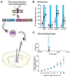
Blue light stimulation of neurons expressing a channelrhodopsin alters cell activity and behavior. (A) A lentiviral vector (LV-CaMKII-ChR2-mCherry) was injected into the motor thalamus of rats to transduce glutamatergic neurons. Blue light stimulation of motor thalamus opened channelrhodopsin2 cation channels in the membrane of transduced neurons. (B) Effect of blue light stimulation of motor thalamus on movement performance in an acute model of parkinsonism. For each pattern, the number of reaches executed by rats is represented for 5 min before, during and after blue light (473 nm) stimulation. Theta burst stimulation (TBS) and the physiological reaching pattern (pReP) are irregular patterns and significantly increased reaching performance. Conversely, the equivalent tonic patterns (15 and 6.2 Hz, respectively) did not improve reaching. p < 0.05, versus prestimulation (Tukey’s test). #p < 0.05 versus pREP pattern (Tukey’s test). Reproduced with permission from Seeger-Armbruster et al. (2015). (C) A similar experiment performed with injection of LV-EF1α-hChR2(H134R)-eYFP into ventrolateral motor thalamus of a rhesus monkey. Upper panel, data show the firing rate (spikes/bin) of one neuron before (-20–0 ms), during (0–1.2 ms), and after (1.2–40 ms) blue light stimulation (1.2 ms pulse). The cell responded with a brief increase of spiking activity just after the pulse of blue light. Bottom panel, the duration of blue light stimulation affected the responses of motor thalamus neurons; longer pulses caused larger increases in firing rate. Reproduced with permission from Galvan et al. (2012).
FIGURE 4
FIGURE 4
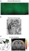
Visualization of lentivirus transduced neurons in post-mortem tissue and in situ imaging. (A) Injection of B19/VSVg-LV-GFP into the lumbar spinal cord resulted in prominent transduction of neurons in the brainstem. Scale bar = 50 μm. Reproduced with permission from Schoderboeck et al. (2015). (B) An electron micrograph of a lentivirus transduced spine in the monkey striatum expressing eYFP. eYFP-tagged neuronal elements were made electron dense by using immunoperoxidase labeling with DAB as the chromogen, prior to general processing for electron microscopy. Reproduced with permission from Galvan et al. (2012). (C) In situ BOLD signals from opto-functional MRI in mice (Desai et al., 2011). Channelrhodopsin-GFP expression following injection of LV-FCK-ChR2- GFP into the somatosensory cortex (left). Voxels with significant increases in BOLD signal (color scale) are shown 1 mm posterior to bregma (right). Reproduced with permission from Desai et al. (2011).

References

    1. Abordo-Adesida E., Follenzi A., Barcia C., Sciascia S., Castro M. G., Naldini L., et al. (2005). Stability of lentiviral vector-mediated transgene expression in the brain in the presence of systemic antivector immune responses. Hum. Gene Ther. 16 741–751 10.1089/hum.2005.16.741 - DOI - PMC - PubMed
    1. Adriani W., Boyer F., Leo D., Canese R., Podo F., Perrone-Capano C., et al. (2010). Social withdrawal and gambling-like profile after lentiviral manipulation of DAT expression in the rat accumbens. Int. J. Neuropsychopharmacol. 13 1329–1342 10.1017/S1461145709991210 - DOI - PubMed
    1. Agwuh K. N., MacGowan A. (2006). Pharmacokinetics and pharmacodynamics of the tetracyclines including glycylcyclines. J. Antimicrob. Chemother. 58 256–265 10.1093/jac/dkl224 - DOI - PubMed
    1. Akerblom M., Sachdeva R., Quintino L., Wettergren E. E., Chapman K. Z., Manfre G., et al. (2013). Visualization and genetic modification of resident brain microglia using lentiviral vectors regulated by microRNA-9. Nat. Commun. 4 1770 10.1038/ncomms2801 - DOI - PubMed
    1. Annoni A., Battaglia M., Follenzi A., Lombardo A., Sergi-Sergi L., Naldini L., et al. (2007). The immune response to lentiviral-delivered transgene is modulated in vivo by transgene-expressing antigen-presenting cells but not by CD4+CD25+ regulatory T cells. Blood 110 1788–1796 10.1182/blood-2006-11-059873 - DOI - PubMed