. 2015 Jun 4;11(6):e1005280.
doi: 10.1371/journal.pgen.1005280. eCollection 2015 Jun.

A legume genetic framework controls infection of nodules by symbiotic and endophytic bacteria

Affiliations

A legume genetic framework controls infection of nodules by symbiotic and endophytic bacteria

Rafal Zgadzaj et al. PLoS Genet. .

Abstract

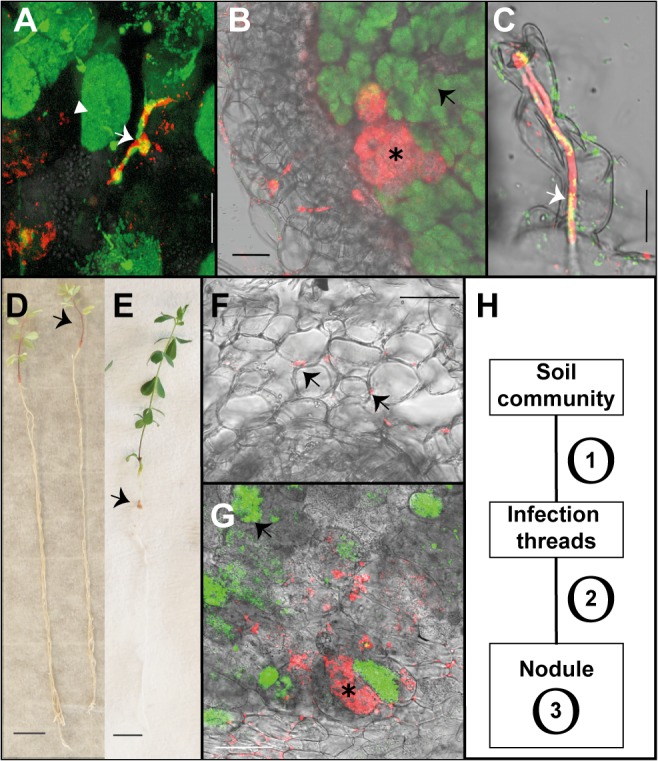
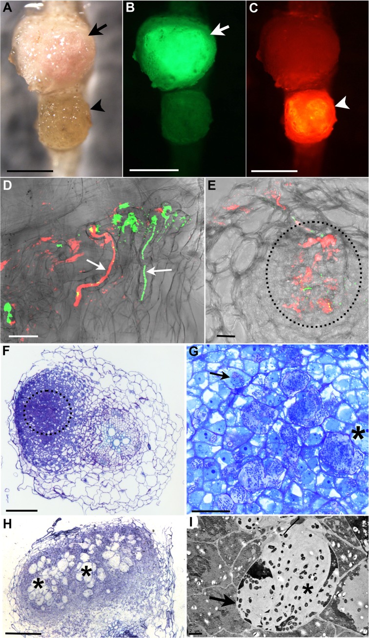
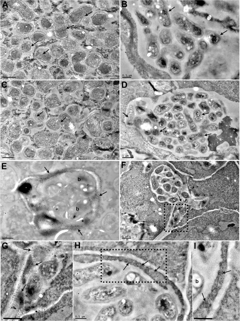
Legumes have an intrinsic capacity to accommodate both symbiotic and endophytic bacteria within root nodules. For the symbionts, a complex genetic mechanism that allows mutual recognition and plant infection has emerged from genetic studies under axenic conditions. In contrast, little is known about the mechanisms controlling the endophytic infection. Here we investigate the contribution of both the host and the symbiotic microbe to endophyte infection and development of mixed colonised nodules in Lotus japonicus. We found that infection threads initiated by Mesorhizobium loti, the natural symbiont of Lotus, can selectively guide endophytic bacteria towards nodule primordia, where competent strains multiply and colonise the nodule together with the nitrogen-fixing symbiotic partner. Further co-inoculation studies with the competent coloniser, Rhizobium mesosinicum strain KAW12, show that endophytic nodule infection depends on functional and efficient M. loti-driven Nod factor signalling. KAW12 exopolysaccharide (EPS) enabled endophyte nodule infection whilst compatible M. loti EPS restricted it. Analysis of plant mutants that control different stages of the symbiotic infection showed that both symbiont and endophyte accommodation within nodules is under host genetic control. This demonstrates that when legume plants are exposed to complex communities they selectively regulate access and accommodation of bacteria occupying this specialized environmental niche, the root nodule.


Conflict of interest statement

The authors have declared that no competing interests exist.

Figures

Fig 1
Fig 1. Endophytic colonisation of Lotus japonicus roots and nodules by R. mesosinicum KAW12.
A) Nodule section displaying a cortical infection thread (arrow) that contains both the M. loti wild-type and KAW12 bacteria, among fully infected nodule cells (arrow head) containing the M.loti symbiont. B) Nodule section showing that KAW12 (*) multiplication inside nodules is limited to small sectors compared to M. loti wild-type (arrow), which is the predominant coloniser. C) Root hair infection thread (arrow) containing both the M. loti wild-type symbiont and the KAW12 endophytic bacteria. D) Lotus plants inoculated with KAW12 endophyte display a nod-minus and nitrogen-starved phenotype (arrow) in comparison to E) Lotus plants that form nodules (arrow) and establish a nitrogen-fixing symbiosis after inoculation with M. loti wild-type. F) Root section illustrating the capacity of KAW12 (arrow) to colonise the intercellular space of Lotus roots. G) Section of an M. loti nodZ-induced nodule presenting KAW12 (*) and M. loti nodZ (arrow) infection. H) The infection and accommodation of compatible endophytes within Lotus nodules is regulated in at least three steps. Scale bars: A) and C) 20 μm, B) F) and G) 50 μm, D) and E) 1 cm. Mesorhizobium loti bacteria are visualized in green, and KAW12 in red. Mixed inoculum has been used in A) to C) and G), and single inocula with the aforementioned bacteria have been used for E) to F).
Fig 2
Fig 2. R. mesosinicum KAW12 colonisation pattern in Lotus japonicus nodules induced by M. loti exoU.
A) to C) Nodules colonised by M. loti exoU-GFP (arrow) or KAW12-DsRed (arrowhead). The two nodules were visualized in bright field (A) with the GFP filter (B) or with the DsRed filter (C). D) Root hair infection threads (arrows) colonised by M. loti exoU (green) and KAW12 (red). E) Confocal laser scanning microscopy (CLSM) image of a nodule section illustrating internal nodule infection (dashed line) by KAW12 (red) and M. loti exoU (green). F) Thin section of a nodule primordium showing KAW12 infection (dashed line) in the inner zone. G) Detailed view of the same nodule as in (F) illustrating the inter- (*) and intra-cellular (arrow) KAW12-containing lagoons. H) Section of a mature nodule presenting multiple and enlarged lagoons colonised by KAW12 (*). (I) Transmission electron micrograph of an intercellular lagoon (arrow) containing bacteria surrounded by a white, undefined matrix (*). Scale bars = 500 μm (A to C), 20 μm (D, G), 50 μm (E), 100 μm (F, H), and 2 μm (I). The M. loti exoU is visualized in green and KAW12 in red (A to E).
Fig 3
Fig 3. Transmission electron micrographs of Lotus japonicus nodules colonised by M. loti exoU or by R. mesosinicum KAW12.
The nodule sections were immunogold labelled (arrows) with an antibody against the GFP protein (A, B), or against the DsRED protein (C, D). GFP was detected (arrows) in the M. loti exoU-selected nodule (A), and DsRED (arrows) in the KAW12-selected nodule (D). There is some minor nonspecific labelling by the GFP antibody (arrows) in the nodule colonised by the DsRed-tagged KAW12 (B) and by the DsRED antibody (arrows) in the nodule colonised by the GFP-tagged M. loti exoU (C). Immunogold labelling of homogalacturonan by the JIM5 monoclonal antibody shows the presence of cell wall material (arrow) in the infection thread that contains M. loti exoU (E), and in the lagoons containing KAW12 (F, G). KAW12 is released inside the plant cell (*) (F). Immunogold labelling of glycoproteins (arrows) by the MAC236 monoclonal antibody reveals their location within the plant cells containing the KAW12-containing lagoons (H, I). Detailed images of the regions marked by rectangles in F) and H) are shown in G) and I), respectively. Scale bars = 1 μm (A to D, and F), 0.5 μm (E, G, I). b = bacteria.

References

    1. Jones JD, Dangl JL. The plant immune system. Nature. 2006. November 16;444(7117):323–9. - PubMed
    1. Kiers ET, Denison RF. Sanctions, Cooperation, and the Stability of Plant-Rhizosphere Mutualisms Annual Review of Ecology Evolution and Systematics. Palo Alto: Annual Reviews; 2008. p. 215–36.
    1. Parniske M. Arbuscular mycorrhiza: the mother of plant root endosymbioses. Nat Rev Microbiol. 2008. October;6(10):763–75. 10.1038/nrmicro1987 - DOI - PubMed
    1. Turner TR, Ramakrishnan K, Walshaw J, Heavens D, Alston M, Swarbreck D, et al. Comparative metatranscriptomics reveals kingdom level changes in the rhizosphere microbiome of plants. The ISME journal. 2013. December;7(12):2248–58. 10.1038/ismej.2013.119 - DOI - PMC - PubMed
    1. Bulgarelli D, Schlaeppi K, Spaepen S, Ver Loren van Themaat E, Schulze-Lefert P. Structure and functions of the bacterial microbiota of plants. Annual review of plant biology. 2013;64:807–38. 10.1146/annurev-arplant-050312-120106 - DOI - PubMed

Publication types

LinkOut - more resources