. 2015 Aug 12;15(8):5321-9.
doi: 10.1021/acs.nanolett.5b01688. Epub 2015 Jun 8.

3D Printed Programmable Release Capsules

Affiliations

3D Printed Programmable Release Capsules

Maneesh K Gupta et al. Nano Lett. .

Abstract

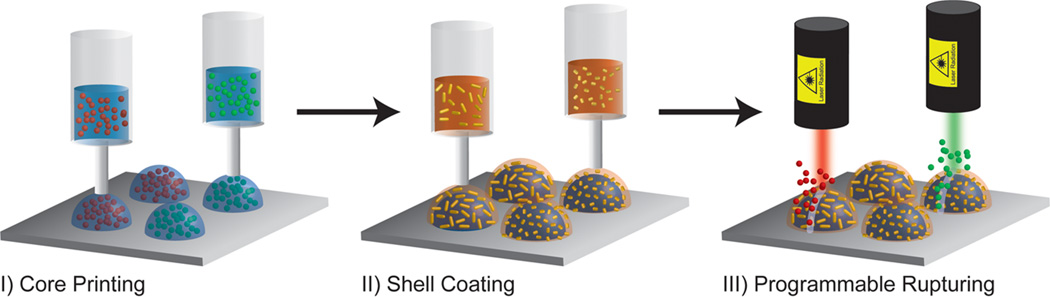
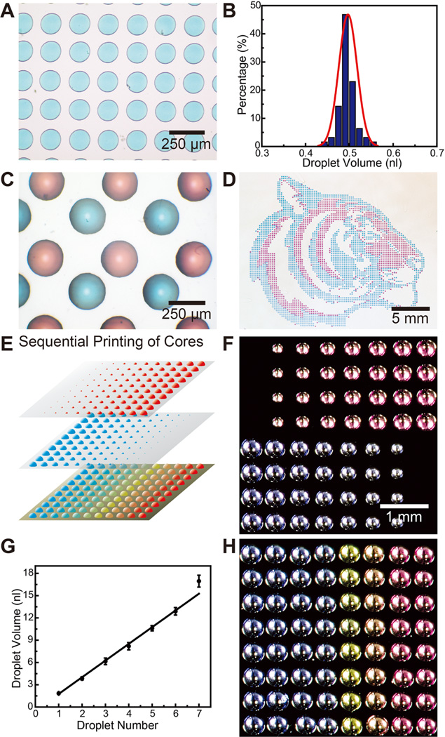
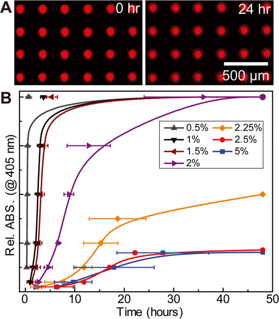
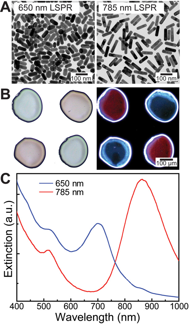
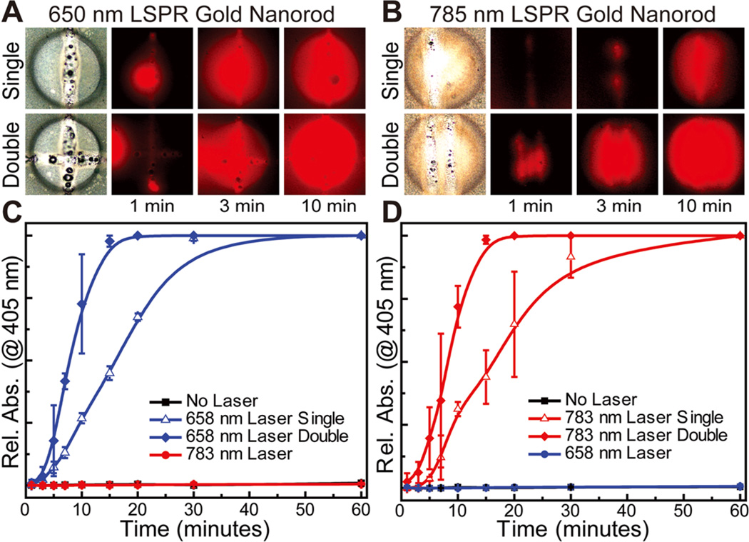
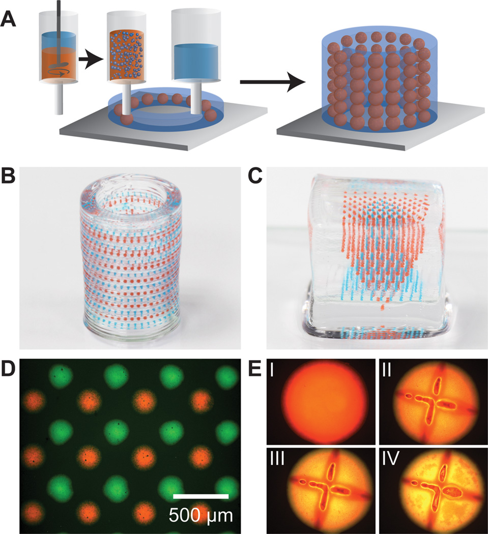
The development of methods for achieving precise spatiotemporal control over chemical and biomolecular gradients could enable significant advances in areas such as synthetic tissue engineering, biotic-abiotic interfaces, and bionanotechnology. Living organisms guide tissue development through highly orchestrated gradients of biomolecules that direct cell growth, migration, and differentiation. While numerous methods have been developed to manipulate and implement biomolecular gradients, integrating gradients into multiplexed, three-dimensional (3D) matrices remains a critical challenge. Here we present a method to 3D print stimuli-responsive core/shell capsules for programmable release of multiplexed gradients within hydrogel matrices. These capsules are composed of an aqueous core, which can be formulated to maintain the activity of payload biomolecules, and a poly(lactic-co-glycolic) acid (PLGA, an FDA approved polymer) shell. Importantly, the shell can be loaded with plasmonic gold nanorods (AuNRs), which permits selective rupturing of the capsule when irradiated with a laser wavelength specifically determined by the lengths of the nanorods. This precise control over space, time, and selectivity allows for the ability to pattern 2D and 3D multiplexed arrays of enzyme-loaded capsules along with tunable laser-triggered rupture and release of active enzymes into a hydrogel ambient. The advantages of this 3D printing-based method include (1) highly monodisperse capsules, (2) efficient encapsulation of biomolecular payloads, (3) precise spatial patterning of capsule arrays, (4) "on the fly" programmable reconfiguration of gradients, and (5) versatility for incorporation in hierarchical architectures. Indeed, 3D printing of programmable release capsules may represent a powerful new tool to enable spatiotemporal control over biomolecular gradients.

Keywords: 3D printing; biomolecular gradients; core−shell particles; plasmonic nanorods; release capsules; spatiotemporal patterning.


Figures

Figure 1
Figure 1
Programmable printing and rupturing of capsules: (I) multiplexed arrays of aqueous cores containing biomolecular payloads are printed directly on a solid substrate; (II) PLGA solutions containing AuNRs of varying lengths are dispensed directly on the aqueous cores, forming a solid stimuli-responsive shell; (III) the capsules are selectively ruptured via irradiation with a laser wavelength corresponding to the absorption peak of the nanorods.
Figure 2
Figure 2
Printing of aqueous cores. (A) Optical micrograph of an array of blue aqueous cores with a center-to-center spacing of 200 µm (colors are from commercial food dyes). (B) The corresponding histogram shows the distribution of droplet volumes. (C) Optical micrograph of a multiplexed array of red and blue capsule cores with spacing of 200 µm between cores of opposite color. (D) Optical image of large scale (>4000) multiplexed patterned red and blue capsule cores in the shape of a “Princeton tiger”. (E) Schematic illustrating the generation of capsule arrays with varying compositions. Arrays with varying volumes are generated by dispensing multiple droplets in the same locations. Overlaying them creates arrays with varying compositions. (F) Optical micrographs of two different pH capsule arrays with varying core volumes. The number of dispensed drops varies from 1 to 8 drops across the array. (G) Plot showing a linear relationship of core volume to the number of droplets dispensed. (H) Optical micrograph of a 2D array with varying pH, adjusted by the ratio of H2SO4 and NaOH within, indicated by the color of m-cresol purple.
Figure 3
Figure 3
Encapsulation of the aqueous core with a PLGA shell. (A) Fluorescent optical micrographs showing the stability of encapsulated cores containing Rhodamine B isothiocyanate–dextran submerged in water over a 24 h period. (B) Plots showing tunable passive release kinetics of HRP from capsule arrays, monitored by an ABTS substrate, achieved by varying the thickness of the PLGA shell (N = 3).
Figure 4
Figure 4
Incorporation of AuNRs in the PLGA shell. (A) TEM images of two different length AuNRs (diameter ~25 nm) with absorption peaks centered at 650 and 785 nm. (B) Bright-field (left) and dark-field (right) optical micrographs of dispensed PLGA shell arrays functionalized with different length nanorods. In the bright-field image, the blue and red colors correspond to the 650 and 780 nm absorption nanorods. The colors are switched in the dark-field image because this shows the scattered component of light as compared to the transmitted in bright-field mode. (C) Vis–NIR spectra of PLGA shells loaded with different length nanorods.
Figure 5
Figure 5
Selective laser-triggered rupture and release of enzymes. (A, B) Bright-field and fluorescent optical images of 650 and 785 nm ruptured capsules showing release of HRP (monitored by Amplex Red), respectively, with release rate controlled by the ruptured area of the PLGA polymer shell (edge length of the square images: 300 µm) (C, D) Plots showing release of HRP from ruptured capsule arrays, demonstrating orthogonal selectivity of AuNR-encrusted capsules as well as fine control over release kinetics, monitored by an ABTS substrate (N = 3).
Figure 6
Figure 6
3D printing of hierarchically multiplexed capsule arrays. (A) Schematic illustrating an emulsion ink-based method to 3D print complex capsule arrays. The emulsion ink is prepared by directly dispersing the aqueous core in the PLGA solution. The hydrogel and emulsion inks are sequentially printed in a layer-by-layer manner to form a 3D structure. (B, C) Optical images of 3D multiplexed capsule arrays directly printed in cylindrical and square hydrogel matrices, respectively (colors of the capsules are from food dyes in the dispersed cores). (D) Fluorescent optical image of a single layer of a multiplexed emulsion-based capsule array. (E) Fluorescent optical images showing rupture and release of fluorescein dye (poly(fluorescein isothiocyanate allylamine hydrochloride)) from an emulsion capsule with Nile red stained PLGA (I: before laser rupture; II, III, IV: 15 min, 1 h, and 2 h after laser rupture; diameter of the capsule: ~300 µm).

References

    1. Biondi M, Ungaro F, Quaglia F, Netti PA. Adv. Drug Delivery Rev. 2008;60:229–242. - PubMed
    1. Griffith LG, Swartz MA. Nat. Rev. Mol. Cell Biol. 2006;7:211–224. - PubMed
    1. Lutolf MP, Gilbert PM, Blau HM. Nature. 2009;462:433–441. - PMC - PubMed
    1. Lutolf MP, Hubbell JA. Nat. Biotechnol. 2005;23:47–55. - PubMed
    1. Kalinin YV, Murali A, Gracias DH. RSC Adv. 2012;2:9707–9726. - PMC - PubMed

Publication types

MeSH terms