. 2015 Jun 4;10(6):e0129907.
doi: 10.1371/journal.pone.0129907. eCollection 2015.

Amyloid properties of the mouse egg zona pellucida

Affiliations

Amyloid properties of the mouse egg zona pellucida

Nathan Egge et al. PLoS One. .

Abstract

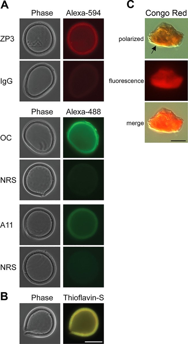
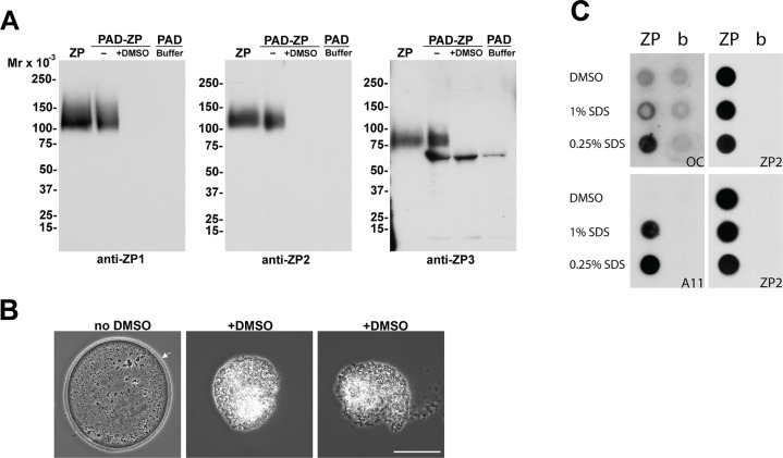
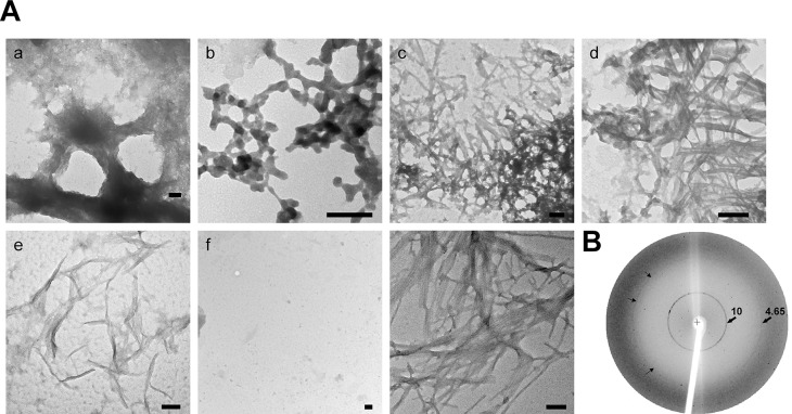
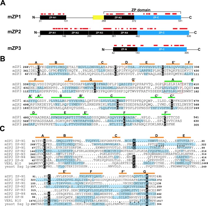
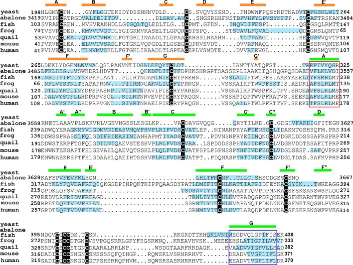
The zona pellucida (ZP) surrounding the oocyte is an extracellular fibrillar matrix that plays critical roles during fertilization including species-specific gamete recognition and protection from polyspermy. The mouse ZP is composed of three proteins, ZP1, ZP2, and ZP3, all of which have a ZP polymerization domain that directs protein fibril formation and assembly into the three-dimensional ZP matrix. Egg coats surrounding oocytes in nonmammalian vertebrates and in invertebrates are also fibrillar matrices and are composed of ZP domain-containing proteins suggesting the basic structure and function of the ZP/egg coat is highly conserved. However, sequence similarity between ZP domains is low across species and thus the mechanism for the conservation of ZP/egg coat structure and its function is not known. Using approaches classically used to identify amyloid including conformation-dependent antibodies and dyes, X-ray diffraction, and negative stain electron microscopy, our studies suggest the mouse ZP is a functional amyloid. Amyloids are cross-β sheet fibrillar structures that, while typically associated with neurodegenerative and prion diseases in mammals, can also carry out functional roles in normal cells without resulting pathology. An analysis of the ZP domain from mouse ZP3 and ZP3 homologs from five additional taxa using the algorithm AmylPred 2 to identify amyloidogenic sites, revealed in all taxa a remarkable conservation of regions that were predicted to form amyloid. This included a conserved amyloidogenic region that localized to a stretch of hydrophobic amino acids previously shown in mouse ZP3 to be essential for fibril assembly. Similarly, a domain in the yeast protein α-agglutinin/Sag 1p, that possesses ZP domain-like features and which is essential for mating, also had sites that were predicted to be amyloidogenic including a hydrophobic stretch that appeared analogous to the critical site in mouse ZP3. Together, these studies suggest that amyloidogenesis may be a conserved mechanism for ZP structure and function across billions of years of evolution.


Conflict of interest statement

Competing Interests: The authors have declared that no competing interests exist.

Figures

Fig 1
Fig 1. Amyloidogenic properties of the mouse ZP.
A) The detection of amyloids in isolated mouse ZP was carried out using the amyloid conformation-dependent antibodies anti-fibrillar OC and anti-oligomer A11 in immunofluorescence analysis. Normal rabbit serum (NRS) served as a control. The anti-ZP3 antibody was used as a marker for the ZP with normal goat IgG serving as a control antibody. The corresponding phase images are shown for each fluorescent image. B) Intact ZP stained with 0.1% thioflavin S to detect amyloids. Scale bar = 50 μm. C) ZP pellets stained with 0.2% Congo Red showed yellow-green birefringence (arrow) when examined under polarizing light and bright red fluorescence when examined with UV light. Scale bar = 10 μm.
Fig 2
Fig 2. Identification of proteins in the ZP amyloid.
A) Thirty isolated ZP were treated with (+) or without (-) DMSO followed by incubation with the PAD ligand and eluted proteins examined by immunoblot using anti-ZP1, ZP2, and ZP3 antibodies. ZP, an equivalent number of ZP placed directly in SDS-PAGE loading buffer without PAD incubation. Buffer, PBS only incubated with PAD beads. Molecular weight markers indicate kDa. B) phase images of oocytes incubated in the presence (+) or absence (no DMSO) of 90% DMSO for 2 minutes at room temperature. Arrow indicates ZP. Scale bar = 50 μm. C) Equal numbers of ZP were exposed to 90% DMSO, 1% SDS or 0.25% SDS prior to spotting on nitrocellulose using a dot blot apparatus. Membranes were then incubated with the amyloid anti-fibrillar OC and anti-oligomer A11 antibodies. Dot blots were rehybridized with anti-ZP2 antibody to confirm the presence of ZP protein in each well. b, buffer.
Fig 3
Fig 3. ZP exhibit structural characteristics of amyloid.
Aa-Ae) Isolated ZP were digested with chymotrypsin followed by spotting on to grids and staining with 2% uranyl acetate. TEM micrographs show the various amyloid-like structures observed in the dispersed ZP. Af) Negative control in which buffer containing chymotrypsin but not ZP was spotted on to a grid. Ag) TEM micrograph of human Aβ amyloid fibrils. Scale Bar = 100 nm. B) X-ray diffraction of mouse ZP. Numbers represent Angstroms. Small arrows indicate the 4.65Å reflection.
Fig 4
Fig 4. Identification of amyloidogenic regions in mouse ZP proteins.
A) Schematic diagram of mature ZP1, ZP2, and ZP3 with amyloidogenic regions predicted by AmylPred 2 indicated as red bars above the individual domains. Yellow box, trefoil domain. Numbers signify amino acid number. B) Structure based sequence alignment of the ZP polymerization domain of mouse ZP1 (aa 268–541), ZP2 (aa 361–630), and ZP3 (aa 42–305) showing amyloidogenic sites (blue highlighting), as predicted by the Amylpred2 algorithm. Cysteine residues are noted by black boxes. The internal hydrophobic patch (IHP) is indicated by a red box [35]. The β-strand secondary structure based on the crystal structure of chicken ZP3 is noted by orange bars (ZP-N subdomain) and green bars (ZP-C subdomain) above the amino acid sequences [8]. C) Structure based alignment of ZP-N domains in mouse ZP1 (N1, N2), ZP2 (N1-N4), ZP3 (N), abalone VERL repeat 10 (R10) and yeast α-agglutinin/Sag 1p showing predicted amyloidogenic sites in blue highlighting as determined by the AmylPred2 algorithm. The β-strand secondary structure, based on structure of mZP-N is indicated by orange bars above the amino acid sequence [7]. Cysteine residues are noted by black boxes. *, indicate sites essential or important for ZP2-sperm and α-agglutinin-a-agglutinin binding [7, 36].
Fig 5
Fig 5. Evolutionary conservation of amyloidogenic sites in the ZP polymerization domain of ZP3 homologs.
Structure based sequence alignment of the ZP polymerization domain of mouse ZP3 and ZP3/egg coat homologs from Haliotis rufescens (red abalone) vitelline envelope receptor of lysin (VERL repeat 23), Oncorhynchus mykiss (rainbow trout) vitelline envelope protein gamma, Xenopus laevis (frog) gp41, Coturnix japonica (Japanese quail) glycoprotein C and human ZP3 with the amyloidogenic sites as predicted by AmylPred2 indicated by blue highlighting. The ZP-N domain in Saccharomyces cerevisiae α-agglutinin/Sag 1 is also shown with its predicted amyloidogenic sites. Cysteine residues are noted by black boxes. The internal hydrophobic patch (IHP) is indicated by a red box and the external hydrophobic patch (EHP) is noted by the blue box [35]. The β-strand secondary structure based on the structure of chicken ZP3 is noted by orange bars (ZP-N subdomain) and green bars (ZP-C subdomain) [8].

References

    1. Bleil JD, Wassarman PM. Synthesis of zona pellucida proteins by denuded and follicle-enclosed mouse oocytes during culture in vitro. Proc Natl Acad Sci USA 1980; 77:1029–1033. - PMC - PubMed
    1. Litscher ES, Qi H, Wassarman PM. Mouse zona pellucida glycoproteins mZP2 and mZP3 undergo carboxy-terminal proteolytic processing in growing oocytes. Biochemistry 1999; 38:12280–12287. - PubMed
    1. Boja ES, Hoodbhoy T, Fales HM, Dean J. Structural characterization of native mouse zona pellucida proteins using mass spectrometry J Biol Chem. 2003; 278:34189–34202. - PubMed
    1. Greve JM, Wassarman PM. Mouse egg extracellular coat is a matrix of interconnected filaments possessing a structural repeat. J Mol Biol. 1985; 181:253–264. - PubMed
    1. Litscher ES, Janssen WG, Darie CC, Wassarman PM. Purified mouse egg zona pellucida glycoproteins polymerize into homomeric fibrils under non-denaturing conditions. J Cell Physiol. 2008; 214:153–157. - PubMed

Publication types

LinkOut - more resources