. 2015 Oct;22(10):822-9.
doi: 10.1038/gt.2015.53. Epub 2015 Jul 2.

CRISPR-Cas9-mediated gene knockout in primary human airway epithelial cells reveals a proinflammatory role for MUC18

Affiliations

CRISPR-Cas9-mediated gene knockout in primary human airway epithelial cells reveals a proinflammatory role for MUC18

H W Chu et al. Gene Ther. 2015 Oct.

Abstract

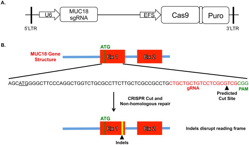
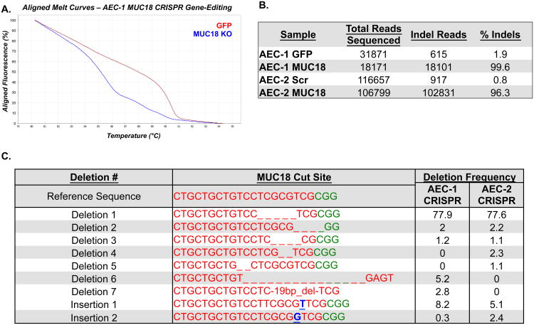
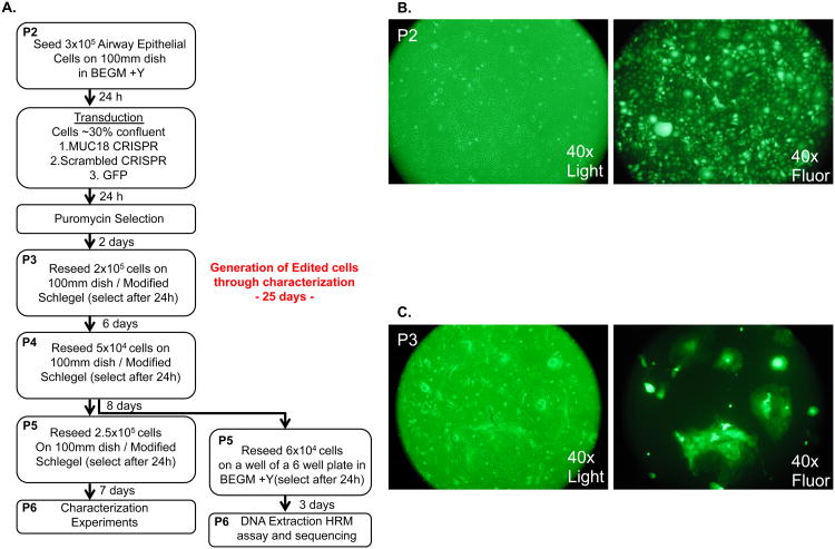
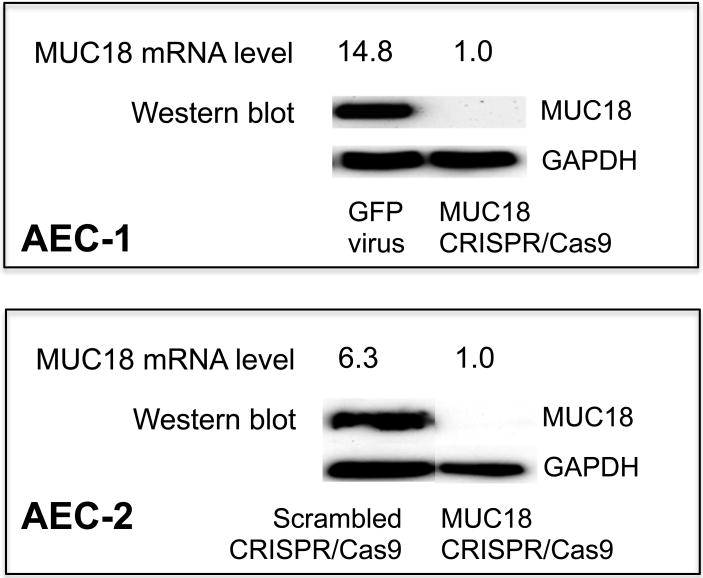
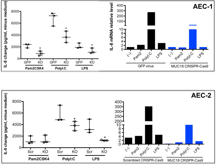
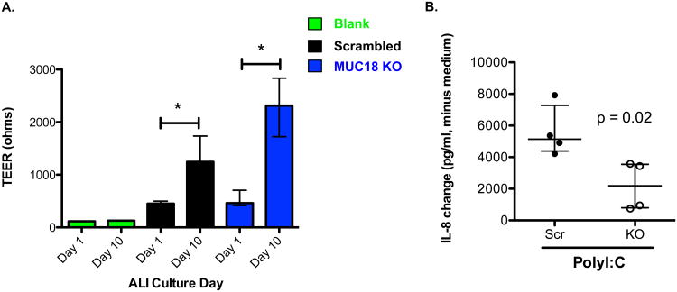
Targeted knockout of genes in primary human cells using CRISPR-Cas9-mediated genome-editing represents a powerful approach to study gene function and to discern molecular mechanisms underlying complex human diseases. We used lentiviral delivery of CRISPR-Cas9 machinery and conditional reprogramming culture methods to knockout the MUC18 gene in human primary nasal airway epithelial cells (AECs). Massively parallel sequencing technology was used to confirm that the genome of essentially all cells in the edited AEC populations contained coding region insertions and deletions (indels). Correspondingly, we found mRNA expression of MUC18 was greatly reduced and protein expression was absent. Characterization of MUC18 knockout cell populations stimulated with TLR2, 3 and 4 agonists revealed that IL-8 (a proinflammatory chemokine) responses of AECs were greatly reduced in the absence of functional MUC18 protein. Our results show the feasibility of CRISPR-Cas9-mediated gene knockouts in AEC culture (both submerged and polarized), and suggest a proinflammatory role for MUC18 in airway epithelial response to bacterial and viral stimuli.


Conflict of interest statement

Conflicts of Interest: The authors have no conflicts of interest to disclose.

Figures

Figure 1
Figure 1. Lentiviral vector and MUC18 Knockout Targeting Strategy
A. Simplified schematic of the published plentiCRISPRv1 vector we used to target MUC18. We designed the MUC18 gRNA and ligated into the plentiCRISPRv1 vector. The plentiCRISPR vector co-expresses both the MUC18 gRNA and the Cas9 protein. B. The gRNA and thus cutting site was chosen to be immediately downstream of the start codon for MUC18 so indel's introduced by non-homologous repair of the double-stranded break would likely disrupt the protein reading frame.
Figure 2
Figure 2. Genomic characterization of MUC18 CRISPR-Cas9 treated and selected AECs
PCR was used to amplify a sequence crossing the MUC18 cut site using genomic DNA isolated from GFP-virus infected, scrambled MUC18 CRISPR-Cas9 virus treated, and MUC18 CRISPR-Cas9 virus treated airway cells in two experiments (different donors). A. High resolution melt (HRM) analysis of PCR amplicons, revealing a significant melting curve shift for the amplicons derived from MUC18 CRISPR-Cas9 treated versus GFP-virus treated AEC-1 cells. B. Results of Next-Generation sequencing analysis of the same PCR amplicons for indels over the MUC18 cut site, revealing the presence of indels in nearly all reads originating from MUC18 CRISPR-Cas9 treated AECs from the two experiments. C. We list the most common indels detected across the MUC18 CRISPR-Cas9 cut site from the two experiments.
Figure 3
Figure 3. Optimized experimental workflow to generate MUC18 knockout primary airway epithelial cells through lentiviral CRISPR-Cas9 treatment
A. We show the time course and culture conditions for generation of AEC-2 MUC18 knockout cells. Reseeding time refers to the MUC18 CRISPR treated cultures, but approximates growth times in scrambled MUC18 CRISPR-Cas9 control cells as well. Note in an extra effort to eliminate potential fibroblast contamination from altering sequencing results, the cells were passed a last time on collagen coated plates in BEGM before DNA isolation. B. Microscopy pictures of P2 cells immediately before passage, revealing high transduction efficiency. C. Microscopy pictures of P3 cells immediately before passage, revealing all epithelial colonies are GFP positive. Note cells are present in colonies because they are being grown with Schlegel culture methods. Also note green tint in the light microscopy picture is due to the GFP filter.
Figure 4
Figure 4. MUC18 mRNA and Protein Expression in MUC18 CRISPR-Cas9 treated AECs
The two MUC18 knockout cell populations (AEC-1 and AEC-2) were cultured under submerged conditions without any TLR agonist stimulation. Cells were harvested for detection of MUC18 mRNA by real-time PCR and MUC18 protein by Western blot.
Figure 5
Figure 5. Pro-inflammatory response of MUC18 knockout AECs to TLR agonist stimulation
The two MUC18 knockout cell populations (AEC-1 and AEC-2) were incubated in triplicate with PBS (medium control) or agonists of TLR2 (Pam2CSK4), TLR3 (polyI:C) and TLR4 (LPS) for 24 hours. Cell supernatants were harvested for detection of IL-8 protein by ELISA (n = 3 replicates for each condition) and IL-8 mRNA by real-time PCR (n=1). IL-8 protein data (median/interquartile range) are expressed as changes of IL-8 in TLR agonist stimulated cells minus IL-8 in medium control cells. *, p< 0.05 comparing KO supernatants to GFP virus or scrambled MUC18 CRISPR-Cas9. Src = scrambled MUC18 CRISPR-Cas9. Pam 2 = Pam2CSK4. KO = MUC18 CRISPR-Cas9.
Figure 6
Figure 6. Air-liquid interface polarized MUC18 knockout AECs havedecreased IL-8 production in response to TLR3 agonist polyI:C
A. MUC18 knockout and scrambled MUC18 CRISPR-Cas9 treated AECs were cultured at air-liquid interface (ALI) for 10 days. Transepithelial electrical resistance(TEER) measurements are shown before and after polarization. Bars represent median and interquartile range for 8 ALI inserts at two different time points. A single empty insert was used to produce the blank measurement at both time points. *, p<0.001. B. Polarized ALI day 10 cells were stimulated in triplicate with polyI:C for 24 hrs. IL-8 protein data (median/interquartile range) are expressed as changes of IL-8 in polyI:C stimulated cells minus IL-8 in medium control cells. Src = scrambled MUC18 CRISPR-Cas9 treated cells. KO = MUC18 knockout cells.

References

    1. Berns K, Hijmans EM, Mullenders J, Brummelkamp TR, Velds A, Heimerikx M, et al. A large-scale RNAi screen in human cells identifies new components of the p53 pathway. Nature. 2004;428(6981):431–7. - PubMed
    1. Jackson AL, Burchard J, Schelter J, Chau BN, Cleary M, Lim L, et al. Widespread siRNA “off-target” transcript silencing mediated by seed region sequence complementarity. Rna. 2006;12(7):1179–87. - PMC - PubMed
    1. Mali P, Yang L, Esvelt KM, Aach J, Guell M, DiCarlo JE, et al. RNA-guided human genome engineering via Cas9. Science. 2013;339(6121):823–6. - PMC - PubMed
    1. Shalem O, Sanjana NE, Hartenian E, Shi X, Scott DA, Mikkelsen TS, et al. Genome-scale CRISPR-Cas9 knockout screening in human cells. Science. 2014;343(6166):84–7. - PMC - PubMed
    1. Wang T, Wei JJ, Sabatini DM, Lander ES. Genetic screens in human cells using the CRISPR-Cas9 system. Science. 2014;343(6166):80–4. - PMC - PubMed

Publication types