Review
. 2015 Jun 5;116(12):1956-70.
doi: 10.1161/CIRCRESAHA.116.304678.

Role of sodium and calcium dysregulation in tachyarrhythmias in sudden cardiac death

Affiliations
Review

Role of sodium and calcium dysregulation in tachyarrhythmias in sudden cardiac death

Stefan Wagner et al. Circ Res. .

Abstract

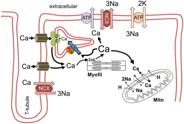
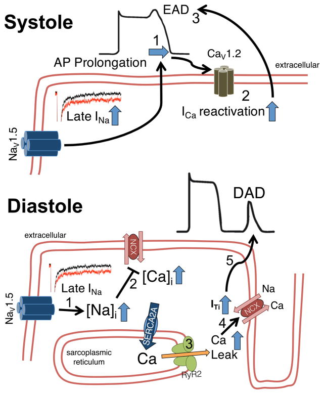
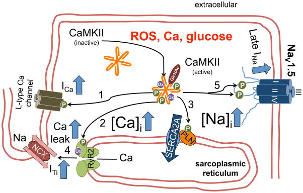
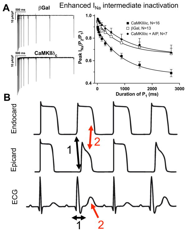
Despite improvements in the therapy of underlying heart disease, sudden cardiac death is a major cause of death worldwide. Disturbed Na and Ca handling is known to be a major predisposing factor for life-threatening tachyarrhythmias. In cardiomyocytes, many ion channels and transporters, including voltage-gated Na and Ca channels, cardiac ryanodine receptors, Na/Ca-exchanger, and SR Ca-ATPase are involved in this regulation. We have learned a lot about the pathophysiological relevance of disturbed ion channel function from monogenetic disorders. Changes in the gating of a single ion channel and the activity of an ion pump suffice to dramatically increase the propensity for arrhythmias even in structurally normal hearts. Nevertheless, patients with heart failure with acquired dysfunction in many ion channels and transporters exhibit profound dysregulation of Na and Ca handling and Ca/calmodulin-dependent protein kinase and are especially prone to arrhythmias. A deeper understanding of the underlying arrhythmic principles is mandatory if we are to improve their outcome. This review addresses basic tachyarrhythmic mechanisms, the underlying ionic mechanisms and the consequences for ion homeostasis, and the situation in complex diseases like heart failure.

Keywords: alternans; arrhythmias; calcium; calcium/calmodulin–dependent kinase II; sodium.


Figures

Figure 1
Figure 1
Schematic diagram of cellular Ca fluxes during excitation-contraction coupling. Ca entry via L-type Ca channels triggers Ca-induced Ca release from the sarcoplasmic reticulum (SR), which results in myofilament activation. For relaxation, cytosolic Ca is transported into the SR via SR Ca-ATPase (SERCA2a) and into the extracellular space via sarcolemmal Na/Ca exchanger. Reproduced with permission from the AHA.
Figure 2
Figure 2
Proarrhythmogenic mechanisms. A) Reentry. Upper panel shows normal conduction around an obstacle. The latter exhibits electrical properties that differ dramatically from the rest of the myocardium (slowed conduction and/or repolarization). The wavefront propagates around this line of block and reaches the back from multiple directions simultaneously resulting in wave annihilation. If, however, unidirectional block (often coupled with decremental conduction) exists around one side (lower panel), the wavefront can reenter and re-excite tissue in front of the unidirectional block. This can lead to stable reentry. B) Triggered activity. Increased depolarizing currents that result in prolongation of action potential duration (APD) can favour L-type Ca channel reactivation. Increased Ca entry via ICa can also lead to SR Ca overload and spontaneous SR Ca release during AP plateau phase. This may generate transient inward (ITi) current by Na/Ca exchange. Both Ca channel reactivation and ITi may depolarize the membrane during AP plateau phase, which can result in an early afterdepolarization (EAD). In addition, SR Ca overload may also result in diastolic SR Ca release. The consequent ITi may lead to depolarization from the resting membrane potential, which can result in a delayed afterdepolarization (DAD). Partially reproduced.
Figure 3
Figure 3
Overview of proarrhythmic consequences of ion channel dysfunction. Congenital or acquired dysfunction of ion channels can result in 1) slowed conduction, 2) repolarization and/or 3) disturbed intracellular Na and Ca homeostatis. Slowed conduction velocity (CV; with or without CV alternans) or repolarization (with or without repolarization alternans) causes spatiotemporal differences in CV and action potential duration (APD) within the myocardium. These regional differences can lead to unidirectional block with reentry and rotor formation. The consequent monomorphic ventricular tachycardia (VT) may result in additional rotors by heart rate-dependent alternans causing polymorphic VT and ventricular fibrillation (VF). Disturbed Na and Ca homeostasis, on the other hand, may lead to Ca alternans, which is linked to repolarization alternans. Moreover, prolongation in APD duration causes EADs and increased Ca leak results in DADs, both of which underlie ectopic activity. The latter can also lead to monomorphic of polymorphic VT and VF.
Figure 4
Figure 4
Characteristics of cardiac alternans. A) Simultaneous recordings of AP and intra-SR free [Ca] in isolated Langendorff-perfused rabbit hearts by optical mapping. At shorter pacing intervals (190 ms), alternans of the AP duration as well as SR Ca release alternans was observed. B) SR Ca release alternans can occur at heart rates where APD alternans is not yet detectable. C) Ca release alternans can be amplified by alternating changes in SR Ca load, i.e. SR Ca load alternans. Data reproduced with permission.
Figure 5
Figure 5
Proarrhythmogenic mechanisms of enhanced late INa. In systole (upper panel), enhanced late INa leads to AP prolongation (1). The longer AP plateau phase increases the likelihood of ICa reactivation (2), which may lead to early afterdepolarizations (EAD, 3). Lower panel: The increased amount of Na influx also results in increased intracellular Na (1), which impairs Ca elimination (2) by the Na/Ca exchanger (either less forward or even increased reverse mode activity). In diastole, the increased intracellular Ca facilitates SR Ca leak (3), which could lead to transient inward current (ITi) by the Na/Ca exchanger (4). The latter can result in delayed afterdepolarizations (DAD, 5).
Figure 6
Figure 6
CaMKII-dependent mechanisms of triggered activity. CaMKII is activated by pathophysiologically relevant stimuli, i.e. increased reactive oxygen species (ROS), increased intracellular Ca, hyperglycemia. CaMKII-dependent phosphorylation of L-type Ca channels (1) may increase ICa window current predisposing to EADs. Increased CaMKII-dependent RyR2 phosphorylation (2) results in increased diastolic Ca leak. CaMKII-dependent phospholamban (PLN) phosphorylation (3) maintains SR Ca content, which may also stimulate diastolic RyR2 openings from the luminal side. Diastolic SR Ca release triggers transient inward current (ITi, 4) and DADs. Increased CaMKII-dependent phosphorylation of NaV1.5 (5) leads to enhanced late INa, which predisposes for both EADs and DADs (Figure 5).
Figure 7
Figure 7
CaMKII-dependent regulation of INa gating and alternans. A) CaMKII-dependent phosphorylation of NaV1.5 has been shown to enhance INa intermediate inactivation (reproduced from Wagner et al with permission; whole-cell patch clamp in rabbit ventricular myocytes). As a result, the number of available Na channels is reduced especially at shorter diastolic intervals. B) Consequences of enhanced INa intermediate inactivation: CV and repolarization alternans. Reduced Na channel availability results in slowed intramural conduction and slowed AP upstroke velocity (1) evident as broader QRS complex on surface ecg (1). In addition, K channel expression is larger in epicardium. Therefore, repolarization is faster in epicardium, especially if Na current is reduced (2). This leads to increased transmural dispersion of repolarization evident as larger and wider T wave on surface ecg (2). Interestingly, Na channels in intermediate inactive state cannot be activated (and become refractory) during the excitation. Thus, these channels are available for the consecutive excitation. Consequently, conduction velocity and AP upstroke velocity will be larger, AP duration longer for the consecutive excitation: the typical pattern of CV and repolarization alternans.

References

    1. Go AS, Mozaffarian D, Roger VL, Benjamin EJ, Berry JD, Blaha MJ, Dai S, Ford ES, Fox CS, Franco S, Fullerton HJ, Gillespie C, Hailpern SM, Heit JA, Howard VJ, Huffman MD, Judd SE, Kissela BM, Kittner SJ, Lackland DT, Lichtman JH, Lisabeth LD, Mackey RH, Magid DJ, Marcus GM, Marelli A, Matchar DB, McGuire DK, Mohler ER, 3rd, Moy CS, Mussolino ME, Neumar RW, Nichol G, Pandey DK, Paynter NP, Reeves MJ, Sorlie PD, Stein J, Towfighi A, Turan TN, Virani SS, Wong ND, Woo D, Turner MB. Heart disease and stroke statistics--2014 update: A report from the american heart association. Circulation. 2014;129:e28–e292. - PMC - PubMed
    1. Lehnart SE, Ackerman MJ, Benson DW, Jr, Brugada R, Clancy CE, Donahue JK, George AL, Jr, Grant AO, Groft SC, January CT, Lathrop DA, Lederer WJ, Makielski JC, Mohler PJ, Moss A, Nerbonne JM, Olson TM, Przywara DA, Towbin JA, Wang LH, Marks AR. Inherited arrhythmias: A national heart, lung, and blood institute and office of rare diseases workshop consensus report about the diagnosis, phenotyping, molecular mechanisms, and therapeutic approaches for primary cardiomyopathies of gene mutations affecting ion channel function. Circulation. 2007;116:2325–2345. - PubMed
    1. Ford ES, Ajani UA, Croft JB, Critchley JA, Labarthe DR, Kottke TE, Giles WH, Capewell S. Explaining the decrease in u.S. Deaths from coronary disease, 1980–2000. N Engl J Med. 2007;356:2388–2398. - PubMed
    1. Dudas K, Lappas G, Stewart S, Rosengren A. Trends in out-of-hospital deaths due to coronary heart disease in sweden (1991 to 2006) Circulation. 2011;123:46–52. - PubMed
    1. Huikuri HV, Castellanos A, Myerburg RJ. Sudden death due to cardiac arrhythmias. N Engl J Med. 2001;345:1473–1482. - PubMed

Publication types

MeSH terms