. 2015 Jun 5:6:7074.
doi: 10.1038/ncomms8074.

TCTEX1D2 mutations underlie Jeune asphyxiating thoracic dystrophy with impaired retrograde intraflagellar transport

Collaborators, Affiliations

TCTEX1D2 mutations underlie Jeune asphyxiating thoracic dystrophy with impaired retrograde intraflagellar transport

Miriam Schmidts et al. Nat Commun. .

Erratum in

Abstract

The analysis of individuals with ciliary chondrodysplasias can shed light on sensitive mechanisms controlling ciliogenesis and cell signalling that are essential to embryonic development and survival. Here we identify TCTEX1D2 mutations causing Jeune asphyxiating thoracic dystrophy with partially penetrant inheritance. Loss of TCTEX1D2 impairs retrograde intraflagellar transport (IFT) in humans and the protist Chlamydomonas, accompanied by destabilization of the retrograde IFT dynein motor. We thus define TCTEX1D2 as an integral component of the evolutionarily conserved retrograde IFT machinery. In complex with several IFT dynein light chains, it is required for correct vertebrate skeletal formation but may be functionally redundant under certain conditions.


Figures

Figure 1
Figure 1. TCTEX1D2 deletion in UCL4 and location of identified variants in TCTEX1D2 protein structure.
(a) The absence of TCTEX1D2 exon 1 and 2 in family UCL4 is visualized by PCR of genomic DNA samples from members of the UCL4 pedigree. TCTEX1D2 exon 4 primers verify the presence of the gene in samples, but TCTEX1D2 exon 2 primers do not amplify in some individuals. Children carrying the homozygous exon 1–2 TCTEX1D2 deletion are marked in black (diagnosed with JATD) or grey (two siblings who were not diagnosed with JATD). The strikethrough indicates death at 2 months of age; double line indicates consanguineous marriage. See also Supplementary Fig. 1. (b) Human TCTEX1D2 (shown above, white boxes indicate untranslated regions (UTR)) consists of five exons encoding a 142 amino-acid protein (shown below) with a C-terminal TCTEX1 domain (blue box). The location of the four identified TCTEX1D2 mutations is shown in the gene (above). Their corresponding location in the protein (below) shows the TCTEX1 domain will be at least partially lost for all variants identified in individuals with JATD.
Figure 2
Figure 2. Clinical features of subjects with TCTEX1D2 mutations.
Affected individuals presented with narrow thorax due to shortened ribs (ac, UCL82 II.1; g, INS family II.1; j, UCL4 II.6), typical pelvis configuration showing trident acetabulum with spurs (arrows) (d, UCL82 II.1; h, INS family II.1; k, UCL4 II.6), polydactyly (e,f, UCL82 II.1; l, UCL4 II.6), shortened extremities (a,b, UCL82 II.1) and brachydactyly (i, INS family II.1, asterisks indicate toes). UCL82 is shown at 38 days in a, c, d and f) and 5.5 yrs in b and e).
Figure 3
Figure 3. Knockdown of tctex1d2 in zebrafish leads to a typical ciliopathy phenotype.
Whole-mount light microscopy showing control morpholino (mo)-injected embryos (ad) and tctex1d2 morphants at 4 days post fertilization (eh). Compared with controls, knockdown of tctex1d2 results in ventrally curved body axis (a,e), small eyes (b,f), pronephric cysts (c,g) and otolith defects (d,h). Alcian blue staining of cartilage identifies craniofacial cartilage defects in tctexd2 morphants (m,n) compared with controls (i,j). Immunofluorescence analysis after staining of cilia at 24 h.p.f. with anti-acetylated tubulin antibody reveals shorter cilia in the pronephric duct of tctex1d2 morphants (o, magnified in p) compared with control embryos (k, magnified in l); however, this difference was no longer evident at 48 h.p.f. (data not shown). Scale bars, 100 μm (aj,m,n) or 50 μm (k,l,o,p).
Figure 4
Figure 4. Loss of human TCTEX1D2 results in retrograde IFT defects.
Immunofluorescence analysis using confocal microscopy revealed an accumulation of IFT88 at the ciliary tips in skin fibroblasts from individual UCL82 II.1 compared with a control (a,b). The accumulation is comparable to that previously reported in fibroblast cilia from an individual with JATD caused by biallelic variants in DYNC2H1 (ref. 34) (c). IFT88 staining is shown in green, anti-acetylated tubulin antibody (red) was used for visualization of the ciliary axoneme, anti-pericentrin antibody (white) marks the ciliary base; the IFT88 labeling also is shown separately in the lower panels. Scale bars, 5 μm. (d) Fraction of cells with IFT88 accumulation at the ciliary tip, 100 cells analysed for each condition. (e) The percentage of ciliated cells in the fibroblast sample from individual UCL82 II.1 compared with control fibroblasts as assessed by counting the number of cilia stained with anti-acetylated tubulin antibody in relation to nuclei stained with DAPI (4,6-diamidino-2-phenylindole, blue in ac) in 10 random visual fields in five independent experiments each, revealing a very mild reduction in ciliation for the TCTEX1D2-deficient cells. One hundred cells were counted per experiment, represented by a single point per experiment. (f) No difference in cilia length between UCL82 II.1 and control fibroblasts was visualized using anti-acetylated tubulin antibody, 100 cells analysed for each condition. Statistical significance in e and f was measured using the Student's t-test, asterisk indicates P value <0.05.
Figure 5
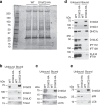
Figure 5. Tctex2b and Tctex1 are in an IFT dynein intermediate chain/light-chain subcomplex with D1bIC2 and D1bIC1.
(a) Flagellar membrane-plus-matrix fractions from wild-type (WT) cells or cells expressing D1bIC2-HA were incubated with anti-HA antibody-conjugated beads. The proteins pulled down by the beads were separated by SDS–PAGE and silver stained. One band (marked *) between 75 and 100 kDa and several bands around 15 kDa (marked ]) are specific for the D1bIC2-HA sample. (bd) Western blots confirming that Tctex2b, Tctex1 and D1bIC1 are specifically co-precipitated with D1bIC2-HA. The unbound and bound samples were probed with the indicated antibodies. In each experiment, all of the D1bIC2-HA was immunoprecipitated from the D1bIC2-HA sample; all of the D1bIC1 (b,d), Tctex1 (b) and Tctex2b (c) was co-precipitated from the D1bIC2-HA sample. None of these proteins were pulled down from the WT control. Some but not all of the DHC1b (d), D1bLIC (b,d), LC7b (c) and LC8 (e) was co-precipitated from the D1bIC2-HA samples; (d) also shows that only very small amounts of the IFT-particle proteins or FLA10 were co-precipitated with D1bIC2-HA. (e) Similar western blot showing that p28 was not co-precipitated with D1bIC2-HA. In b, d and e, the ratio of unbound: bound protein loaded was 1:4; in c, the ratio was 1:2. All antibodies used for Chlamydomonas protein analysis are listed in Supplementary Table 6.
Figure 6
Figure 6. Loss of Tctex2b causes IFT dynein instability and a retrograde IFT defect in Chlamydomonas.
(a) The tctex2b mutant has defects in flagella regeneration. The tctex2b null mutant and A54-e18 (the wild-type strain from which tctex2b was derived) were deflagellated and then allowed to regrow their flagella. Flagella lengths were measured before deflagellation and at time points after deflagellation. The two strains had identical flagellar lengths (11.4 μm) before deflagellation. For each time point, one flagellum from each of 50 cells was measured; error bars are s.d. (b) The tctex2b mutant is defective in retrograde IFT. IFT was recorded in wild-type (A54-e18) and tctex2b flagella by DIC microscopy, and kymograms generated from the video recordings. Tracks with positive slopes represent IFT particles moving anterogradely, and tracks with negative slopes represent particles moving retrogradely. Compared with wild type, few retrograde tracks are visible in the tctex2b kymogram, and these had a much reduced slope. Retrograde particles had a larger apparent size in mutants; similar findings were reported for a temperature-sensitive dhc1b mutant. (c) Quantitative analysis of IFT in wild type (A54-e18) and tctex2b. In tctex2b, anterograde IFT velocity is about the same as in wild type, while anterograde frequency is only slightly reduced, but both retrograde IFT velocity and frequency are greatly reduced. n, number of flagella analysed. Error bars show s.d. (d) Western blot showing reduced IFT dynein subunits in tctex2b whole-cell lysates. Wild-type (A54-e18) and tctex2b whole-cell lysates were probed with antibodies to IFT dynein subunits and IFT-particle proteins. DHC1b, D1bIC2 and D1bLIC are reduced in tctex2b whole-cell lysate. No significant changes were detected for IFT proteins. The same samples were probed for tubulin as loading control. (e) IFT dynein is greatly reduced in tctex2b flagella. Wild-type (A54-e18) and tctex2b flagella were probed with antibodies to IFT dynein subunits and IFT-particle proteins. IFT dynein subunits DHC1b, D1bIC2 and D1bLIC are greatly reduced in tctex2b flagella. IFT-A protein IFT139 and IFT-B proteins IFT172 and IFT81 are increased in tctex2b flagella, consistent with a retrograde IFT defect. The same samples were probed for tubulin as loading control.
Figure 7
Figure 7. Proposed model of IFT dynein composition in Homo sapiens (Hs) and C. reinhardtii (Cr).
Left, IFT dynein (dynein 2/1b) is composed of dynein heavy chains (DYNC2H1(Hs)/DHC1b(Cr); shown in dark grey), dynein light-intermediate chains (DYNC2LI1(Hs)/D1bLIC(Cr); shown in light grey) and different dynein intermediate and light chains (coloured, shown in detail on the right). Right, dynein intermediate chains (WDR34(Hs)/D1bIC2(Cr) and WDR60(Hs)/D1bIC1(Cr)) interact with different dynein light-chain subtypes, including TCTEX1D2(Hs)/TCTEX2b(Cr). Question marks indicate LC7a and DYNLRB2 as unconfirmed components suggested from our Chlamydomonas and human results.

References

    1. Fliegauf M., Benzing T. & Omran H. When cilia go bad: cilia defects and ciliopathies. Nat. Rev. Mol. Cell. Biol. 8, 880–893 (2007) . - PubMed
    1. Hildebrandt F., Benzing T. & Katsanis N. Ciliopathies. N. Engl. J. Med. 364, 1533–1543 (2011) . - PMC - PubMed
    1. Pedersen L.B. & Rosenbaum J.L. Intraflagellar transport (IFT) role in ciliary assembly, resorption and signalling. Curr. Topics Dev. Biol. 85, 23–61 (2008) . - PubMed
    1. Patel-King R.S., Gilberti R.M., Hom E.F. & King S.M. WD60/FAP163 is a dynein intermediate chain required for retrograde intraflagellar transport in cilia. Mol. Biol. Cell 24, 2668–2677 (2013) . - PMC - PubMed
    1. Hom E.F.Y. et al.. A unified taxonomy for ciliary dyneins. Cytoskeleton (Hoboken NJ) 68, 555–565 (2011) . - PMC - PubMed

Publication types

Supplementary concepts