. 2015 Jun 5;348(6239):1143-7.
doi: 10.1126/science.aaa9529.

Human oocytes. Error-prone chromosome-mediated spindle assembly favors chromosome segregation defects in human oocytes

Affiliations

Human oocytes. Error-prone chromosome-mediated spindle assembly favors chromosome segregation defects in human oocytes

Zuzana Holubcová et al. Science. .

Abstract

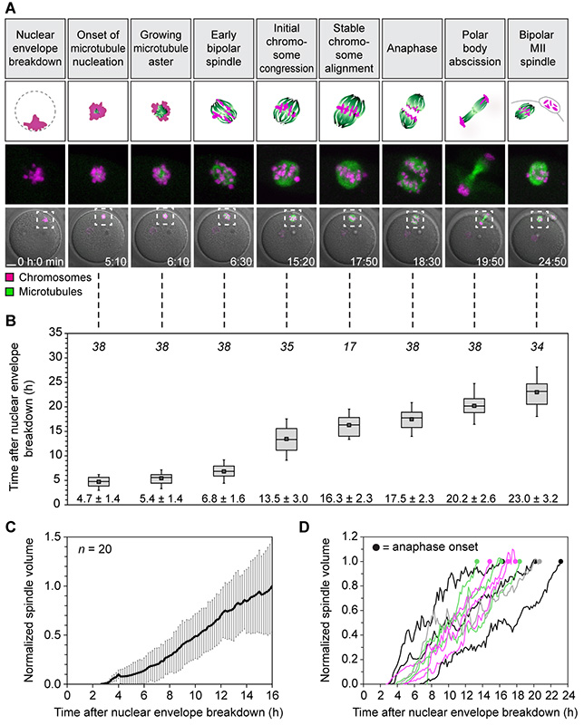
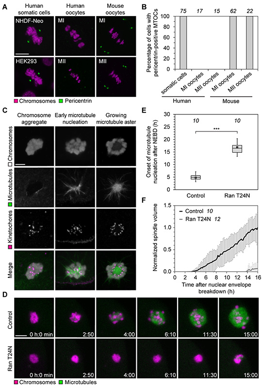
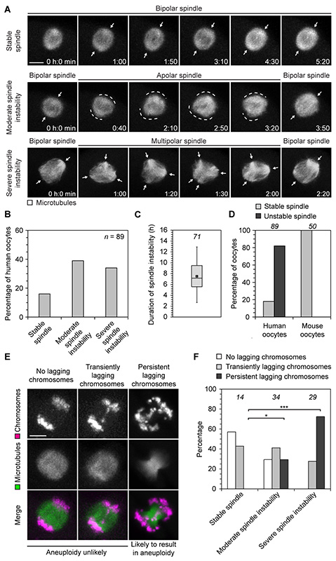
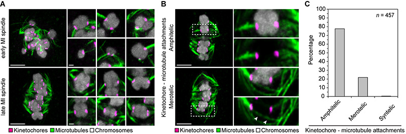
Aneuploidy in human eggs is the leading cause of pregnancy loss and several genetic disorders such as Down syndrome. Most aneuploidy results from chromosome segregation errors during the meiotic divisions of an oocyte, the egg's progenitor cell. The basis for particularly error-prone chromosome segregation in human oocytes is not known. We analyzed meiosis in more than 100 live human oocytes and identified an error-prone chromosome-mediated spindle assembly mechanism as a major contributor to chromosome segregation defects. Human oocytes assembled a meiotic spindle independently of either centrosomes or other microtubule organizing centers. Instead, spindle assembly was mediated by chromosomes and the small guanosine triphosphatase Ran in a process requiring ~16 hours. This unusually long spindle assembly period was marked by intrinsic spindle instability and abnormal kinetochore-microtubule attachments, which favor chromosome segregation errors and provide a possible explanation for high rates of aneuploidy in human eggs.


Figures

Fig. 1
Fig. 1. Stages of meiosis in live human oocytes
(A) Stages of meiosis in human oocytes determined from live human oocytes expressing EGFP-MAP4 (microtubules) and H2B-mRFP1 (chromosomes). A schematic representation of each stage (scheme; microtubules in green; chromosomes in magenta) and stage-specific time-lapse images (z-projections, 4 sections, every 5 μm) merged with differential interference contrast [DIC] are shown (bottom row). Outlined regions are magnified above (middle row). Scale bar, 20 μm. Time displayed in hours: minutes. (B) Quantification of timing of meiotic progression from live oocytes expressing EGFP-MAP4 (microtubules) and H2B-mRFP1 (chromosomes) as shown in (A). The box plot shows median (line), mean (small square), and 25th and 75th (boxes), 5th and 95th percentile (whiskers) of time after NEBD. The number of oocytes is specified in italics. Only oocytes in which the whole maturation process was recorded (from before NEBD to bipolar MII spindle formation) were included. (C and D) The spindle volume was quantified in live human oocytes expressing EGFP-MAP4 (microtubules) as shown in (A). Averaged data from 20 oocytes (C) and examples of individual curves up until anaphase onset (D) are shown.
Fig. 2
Fig. 2. Chromosomes mediate spindle assembly in human oocytes
(A) Immunofluorescence staining of pericentrin and chromosomes (Hoechst) in somatic cells, mouse and human MI and MII oocytes. Scale bars, 10 μm. (B) Spindles of somatic cells as well as metaphase I (MI) and metaphase II (MII) spindles in mouse and human oocytes as shown in (A) were scored for the presence of pericentrin-positive MTOCs. The number of cells is specified in italics. (C) Immunofluorescence staining (z-projections of 6 sections, every 0.3 μm) of kinetochores (CREST), microtubules (α-tubulin) and chromosomes (Hoechst) in human oocytes fixed at different times shortly after NEBD. Scale bar, 10 μm. (D) Live human oocytes expressing H2B-mRFP1 (chromosomes) and EGFP-MAP4 (microtubules) upon microinjection with Ran T24N (lower panel) or BSA (top panel). z-projections of 4 sections, every 5 μm. Scale bar, 10 μm. (E) Onset of microtubule nucleation in live human oocytes expressing EGFP-MAP4 upon microinjection with either Ran T24N or BSA. Box plot as in Fig. 1B. The number of oocytes is specified in italic. ***P<10−14 (t-test). Two Ran T24N-injected oocytes never nucleated microtubules. (F) The spindle volume was quantified in live human oocytes expressing EGFP-MAP4 upon microinjection with either Ran T24N or BSA. The number of oocytes is specified in italics.
Fig. 3
Fig. 3. Spindle instability correlates with chromosome segregation errors
(A) Live human oocytes expressing EGFP-MAP4 (microtubules). z-projections of 4-5 sections, every 3-4 μm. Arrows highlight defined spindle poles; dashed lines mark undefined spindle poles. Scale bar, 10 μm. Polar body extrusion in these cells is shown in fig. S5B. (B) Live human oocytes expressing EGFP-MAP4 as shown in (A) were scored for the presence and degree of spindle instability. n specifies number of oocytes. (C) The duration of spindle instability was measured in live human oocytes expressing EGFP-MAP4 as shown in (A). Box plot as in Fig. 1B. The number of oocytes is specified in italics. (D) Live human and mouse oocytes expressing EGFP-MAP4 (microtubules) were scored for the presence of spindle instability. The number of oocytes is specified in italic. (E) Illustration of classes of lagging chromosomes in live human oocytes expressing H2B-mRFP1 (chromosomes) and EGFP-MAP4 (microtubules). z-projections of 3-5 sections, every 3-5 μm. Scale bar, 10 μm. (F) Live human oocytes expressing H2B-mRFP1 (chromosomes) and EGFP-MAP4 (microtubules) as shown in (E) were scored for the presence of transiently lagging or persistent lagging chromosomes. The number of oocytes is specified in italics. *P<0.05, ***P<10−6 (Fisher’s exact test).
Fig. 4
Fig. 4. Correction of kinetochore-microtubule attachments is incomplete close to anaphase
(A) Immunofluorescence staining (z-projections of 13 sections, every 0.3 μm) of kinetochores (CREST) and microtubules (α-tubulin) in cold-treated human oocytes fixed during early or late spindle assembly. Chromosomes labelled with Hoechst. Scale bar overview, 5 μm. High resolution images of 6 individual chromosome bivalents from overview are shown on the right (z-projections of 2 sections, every 0.3 μm). Scale bar details, 1 μm. (B) Illustration of amphitelic and merotelic kinetochore-microtubule attachments. Immunofluorescence staining (z-projections of 3 sections, every 0.3 μm) of kinetochores (CREST) and microtubules (α-tubulin) in cold-treated human oocytes fixed close to anaphase onset. Chromosomes labelled with Hoechst. The outlined regions are magnified on the right. Arrowheads highlight merotelically attached microtubules. Scale bar, 5 μm. (A and B) All images were deconvolved. Background signal outside of the spindle area was masked in kinetochore channel (magenta). (C) 10 cold-treated oocytes fixed close to anaphase onset (as shown in B) were scored for amphitelic, merotelic or syntelic kinetochore-microtubule attachments. In one case, the bivalent’s kinetochores were attached to the same instead of opposite poles (syntelic attachment).

References

    1. Nagaoka SI, Hassold TJ, Hunt PA. Human aneuploidy: mechanisms and new insights into an age-old problem. Nat Rev Genet. 2012;13:493–504. - PMC - PubMed
    1. Knouse KA, Wu J, Whittaker CA, Amon A. Single cell sequencing reveals low levels of aneuploidy across mammalian tissues. Proc Natl Acad Sci U S A. 2014;111:13409–13414. - PMC - PubMed
    1. Pacchierotti F, Adler ID, Eichenlaub-Ritter U, Mailhes JB. Gender effects on the incidence of aneuploidy in mammalian germ cells. Environ Res. 2007;104:46–69. - PubMed
    1. Templado C, Vidal F, Estop A. Aneuploidy in human spermatozoa. Cytogenet Genome Res. 2011;133:91–99. - PubMed
    1. Danylevska A, Kovacovicova K, Awadova T, Anger M. The frequency of precocious segregation of sister chromatids in mouse female meiosis I is affected by genetic background. Chromosome Res. 2014;22:365–373. - PubMed

Publication types

Substances