Case Reports
. 2015 Jun 4:2015:bcr2015209739.
doi: 10.1136/bcr-2015-209739.

Ameloblastic fibro-odontoma

Affiliations
Case Reports

Ameloblastic fibro-odontoma

Ramlal Gantala et al. BMJ Case Rep. .

Abstract

Ameloblastic fibro-odontoma is a slow growing, benign, expansile epithelial odontogenic tumour with odontogenic mesenchyme, accounting for 0.3-1.7% of jaw tumours, signifying its rarity. The WHO defines it as "a neoplasm composed of proliferating odontogenic epithelium in a cellular ectomesenchymal tissue with varying degrees of inductive changes and dental hard tissue formation". We report a case of an 11-year-old girl who presented to the Department of Maxillo-Facial Medicine and Radiology for the evaluation of a swelling in the left posterior mandible. Her clinical chart and investigations unveiled it as ameloblastic fibro-odontoma. After a promising presurgical evaluation, the lesion was enucleated using an intraoral approach followed by osteoplasty. Osteogenesis was attained despite of any definitive techniques to promote bone regeneration. Immediate postoperative inter-maxillary fixation was performed to prevent pathological fractures for a period of 3 weeks. In an 8-month follow-up, no untoward complications were noticed.


Figures

Figure 1
Figure 1
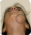
Extraoral picture depicting an ill-defined oval-shaped swelling on the left posterior body of the mandible altering the normal anatomic contour of the lower border.
Figure 2
Figure 2
Clinical presentation shows an ill-defined, diffuse swelling on the left posterior body of the mandible with bi-cortical expansion.
Figure 3
Figure 3
Periapical view with respect to 75 and 36 reports a well-defined homogenous radiopaque mass distal to 75.
Figure 4
Figure 4
Orthopantomogram showing mixed dentition status with a radiopaque mass distal to 75 radiated by radiolucent and sclerotic borders. The displacement of inferior alveolar nerve and altered contour of the lower border of the mandible can be appreciated.
Figurec 5
Figurec 5
(A) CT–axial section revealing expanded cortices with a hyperdense area surrounded by hypodense rim in the left body of the mandible. (B) Three-dimensional (3D) reconstucted image illustrating altered lower border of the mandible with thin lingual cortex. (C): 3D-reconstruted sagittal view presenting hyperdense area surrounded by a hypodense rim (suggestive of fibrous-capsule) inferior to which is the impacted tooth with varying radiodensities of the hyperdense mass and tooth proper.
Figure 6
Figure 6
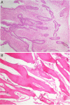
(A) Photomicrograph showing islands and strands of odontogenic epithelium lined by tall columnar cells resembling ameloblasts and central stellate reticulum-like tissue scattered within a highly cellular connective tissue resembling primitive dental papilla. (B) Decalcified H&E stained section revealing odontoma component with dentin containing dentinal tubules, scalloped dentinoenamel junction, enamel space and tissue resembling pulp.
Figure 7
Figure 7
(A) Surgical session showing the approach gained intraorally. (B) Intraoperative photograph after enucleation of the lesion. (C) Enucleated tumour along with the impacted 36.
Figure 8
Figure 8
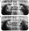
(A) Postoperative orthopantomogram. (B): Follow-up orthopantomogram after 8 months.
Figure 9
Figure 9
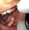
Follow-up intraoral picture after 8 months.

References

    1. Gogri AA, Kadam SG, Umarji HR et al. . Ameloblastic fibro-odontoma differentiating into odontoma: an old concept revised. J Indian Acad Oral Med Radiol 2014;26:310–14. 10.4103/0972-1363.145016 - DOI
    1. De Riu G, Meloni SM, Contini M et al. . Ameloblastic fibro-odontoma. Case report and review of literature. J Craniomaxillofac Surg 2010;38:141–4. 10.1016/j.jcms.2009.04.009 - DOI - PubMed
    1. Pontes HA, Pontes FS, Lameira AG et al. . Report of four cases of ameloblastic fibro-odontoma in mandible and discussion of the literature about the treatment. J Craniomaxillofac Surg 2012;40:e59–63. 10.1016/j.jcms.2011.03.020 - DOI - PubMed
    1. Wood NK, Goaz PW, Kallal R. Mixed radiolucent–radiopaque lesions associated with teeth in differential diagnosis of oral and maxillofacial lesions. 5th edn Elsevier 2006:423–7.
    1. Surej Kumar LK, Manuel S, Khalam SA et al. . Ameloblastic fibro-odontoma. Int J Surg Case Rep 2014;5:1142–4. 10.1016/j.ijscr.2014.11.025 - DOI - PMC - PubMed

Publication types