. 2015 Jul 17;290(29):17935-17945.
doi: 10.1074/jbc.M115.665513. Epub 2015 Jun 4.

Structural Basis of Clade-specific Engagement of SAMHD1 (Sterile α Motif and Histidine/Aspartate-containing Protein 1) Restriction Factors by Lentiviral Viral Protein X (Vpx) Virulence Factors

Affiliations

Structural Basis of Clade-specific Engagement of SAMHD1 (Sterile α Motif and Histidine/Aspartate-containing Protein 1) Restriction Factors by Lentiviral Viral Protein X (Vpx) Virulence Factors

Ying Wu et al. J Biol Chem. .

Abstract

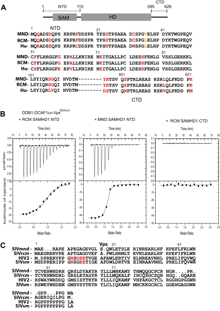
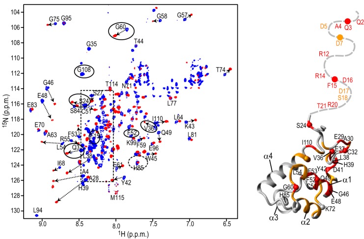
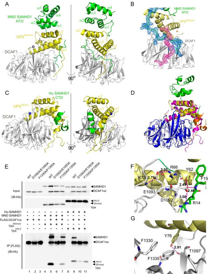
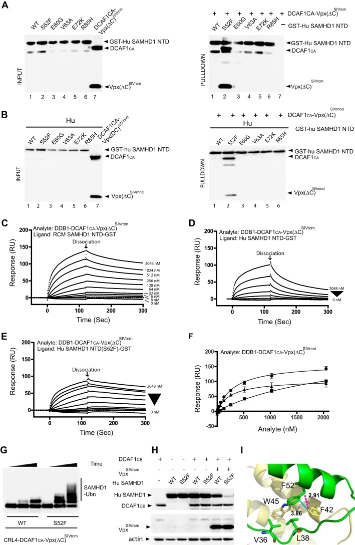
Sterile α motif (SAM) and histidine/aspartate (HD)-containing protein 1 (SAMHD1) restricts human/simian immunodeficiency virus infection in certain cell types and is counteracted by the virulence factor Vpx. Current evidence indicates that Vpx recruits SAMHD1 to the Cullin4-Ring Finger E3 ubiquitin ligase (CRL4) by facilitating an interaction between SAMHD1 and the substrate receptor DDB1- and Cullin4-associated factor 1 (DCAF1), thereby targeting SAMHD1 for proteasome-dependent down-regulation. Host-pathogen co-evolution and positive selection at the interfaces of host-pathogen complexes are associated with sequence divergence and varying functional consequences. Two alternative interaction interfaces are used by SAMHD1 and Vpx: the SAMHD1 N-terminal tail and the adjacent SAM domain or the C-terminal tail proceeding the HD domain are targeted by different Vpx variants in a unique fashion. In contrast, the C-terminal WD40 domain of DCAF1 interfaces similarly with the two above complexes. Comprehensive biochemical and structural biology approaches permitted us to delineate details of clade-specific recognition of SAMHD1 by lentiviral Vpx proteins. We show that not only the SAM domain but also the N-terminal tail engages in the DCAF1-Vpx interaction. Furthermore, we show that changing the single Ser-52 in human SAMHD1 to Phe, the residue found in SAMHD1 of Red-capped monkey and Mandrill, allows it to be recognized by Vpx proteins of simian viruses infecting those primate species, which normally does not target wild type human SAMHD1 for degradation.

Keywords: SAM domain and HD domain-containing protein 1 (SAMHD1); SIV; X-ray crystallography; human immunodeficiency virus (HIV); nuclear magnetic resonance (NMR); restriction factor; ubiquitylation (ubiquitination).


Figures

FIGURE 1.
FIGURE 1.
Vpx from SIVrcm (VpxSIVrcm) binds to the N terminus of RCM and MND SAMHD1 and recruits SAMHD1 to DDB1-DCAF1. A, schematic representation of the SAMHD1 structure and amino acid sequence alignment of the NTD (residues 1–115) and the CTD (residues 595–626) of MND, RCM, and Hu SAMHD1. Amino acid residues that vary between monkey and human SAMHD1 are colored in red, and the single residue that differs between MND and RCM SAMHD1 is colored in orange. B, isothermal titration calorimetry traces for RCM SAMHD1 NTD, MND SAMHD1 NTD, and RCM SAMHD1 CTD binding to DDB1-DCAF1CA-VpxSIVrcm. Aliquots of RCM SAMHD1 NTD (30 μm) were added to the DDB1-DCAF1CA-VpxSIVrcm complex (4.6 μm) in a buffer containing 20 mm HEPES, pH 7.4, 150 mm NaCl, and 0.02% sodium azide at 12 °C (left panel). Aliquots of MND SAMHD1 NTD (30 μm) and RCM SAMHD1 CTD (residues 595–626 at 30 μm) were added to the DDB1-DCAF1CA-VpxSIVrcm complex (2 μm) in the same buffer, respectively (middle panel and right panel). The heat of dilution at each concentration and buffer condition was determined and subtracted. The data in the left panel and middle panel were best fitted to a single-site binding isotherm yielding a Kd of 170 ± 22 nm and 22 ± 2 nm, respectively. C, amino sequence alignment of VpxSIVmnd, VpxSIVrcm, VpxHIV-2, and VpxSIVsm. The sequence motif essential for SAMHD1 C-terminal binding is colored in red (29, 30).
FIGURE 2.
FIGURE 2.
Structural mapping of the DCAF1-VpxSIVmnd interaction onto the SAM domain. A Superposition of the 900-MHz 1H,15N TROSY-HSQC spectra of the MND SAMHD1 NTD (52 μm) in the absence (blue) and presence (red) of 52 μm DCAF1-VpxSIVmnd at 298 K (left panel). Selected assignments are provided for several representative, well separated resonances, labeled on free MND SAMHD1 NTD spectrum. Resonances in DCAF1-VpxSIVmnd-bound state are tentatively assigned and connected to the free resonances by arrows. Resonances of the residues that are different in monkey and human are enclosed by ovals. The random coil region of the spectrum is shown by a dashed rectangle. Structural mapping of residues that exhibit large 1H,15N chemical shift perturbations upon DCAF1-VpxSIVmnd binding is provided (right panel). Residues that experience perturbations >0.15 ppm and between 0.07 and 0.15 ppm are colored in red and orange, respectively, onto the backbone structure of the human NTD region (residues 23–118; PDB code 2E8O). The N-terminal flexible tail (residues Met-1–Pro22) for which no coordinates are available is represented by a gray dashed line.
FIGURE 3.
FIGURE 3.
Complex formation between DCAF1-VpxSIVmnd and MND SAMHD1. A, crystal structures of the DCAF1-VpxSIVmnd-MND SAMHD1 NTD complex. All proteins are displayed in ribbon representation. DCAF1, VpxSIVmnd, and MND SAMHD1 NTD are colored in light gray, yellow, and green, respectively. B, electron density for residues Lys-11–Glu-30 of MND SAMHD1 NTD and residues Glu-7–Leu-17 of VpxSIVmnd, contoured at 1σ. C, crystal structures of the DCAF1-VpxSIVsm-Hu SAMHD1 CTD complexes (29). DCAF1, VpxSIVsm, and Hu SAMHD1 CTD are colored in light gray, yellow, and green, respectively. D, superposition of the structures of DCAF1-VpxSIVmnd-MND SAMHD1 NTD and DCAF1-VpxSIVsm-Hu SAMHD1 CTD. In the DCAF1-VpxSIVmnd-MND SAMHD1 NTD complex, the individual components DCAF1, VpxSIVmnd, and MND SAMHD1 NTD are colored in light gray, yellow, and green, respectively. In the DCAF1-VpxSIVsm-Hu SAMHD1 CTD complex, the individual components DCAF1, VpxSIVsm, and Hu SAMHD1 CTD are colored in blue, magenta, and orange, respectively. E, immunoprecipitation (IP) of complexes. FLAG-tagged WT or mutant DCAF1 proteins (residues 1040–1400) were transiently expressed in the presence of MND SAMHD1 and VpxSIVmnd or Hu SAMHD1 and VpxHIV-2 as indicated. Cells were treated with MG132 for 6 h before cell lysates were subjected to immunoprecipitation with anti-FLAG antibody and analyzed by immunoblotting (IB). F and G, hydrogen bonds formed by DCAF1 residues Asp-1092/Glu-1093 (F) and hydrophobic interactions involving DCAF1 residues Phe-1330/Phe-1355 (G) in the DCAF1-VpxSIVmnd-MND SAMHD1 NTD complex. DCAF1, VpxSIVsm, and Hu SAMHD1 NTD are colored in light gray, yellow, and green, respectively.
FIGURE 4.
FIGURE 4.
A single residue in the SAM domain of human SAMHD1 is critical for binding to DDB1-DCAF1-VpxSIVrcm and DDB1-DCAF1-VpxSIVmnd. A and B, GST-pulldown assay of GST-Hu SAMHD1 NTD interacting with DDB1-DCAF1CA-Vpx(ΔC)SIVrcm (A) and DDB1-DCAF1CA-Vpx(ΔC)SIVmnd (B). Hu SAM domain residues that differ from the simian protein were individually changed to those present in MND SAMHD1 and RCM SAMHD1 (see Fig. 1A). Pulldown assays of DDB1-DCAF1CA-Vpx(ΔC)SIVrcm or DDB1-DCAF1CA-Vpx(ΔC)SIVmnd protein complexes with GST-Hu SAMHD1 NTD were immunoblotted with the appropriate antibodies. C–E, real-time kinetic analysis DDB1-DCAF1-VpxSIVrcm binding to NTD. SPR sensorgrams of DDB1-DCAF1CA-Vpx(ΔC)SIVrcm binding to RCM SAMHD1 NTD-GST (C), Hu SAMHD1 NTD-GST (D), and Hu SAMHD1 NTD(S52F)-GST (E). The concentrations of DDB1-DCAF1CA-Vpx(ΔC)SIVrcm were 8, 16, 32, 64, 128, 256, 512, 1024, and 2048 nm. F, binding isotherms of DDB1-DCAF1CA-Vpx(ΔC)SIVrcm binding to RCM SAMHD1 NTD-GST (solid circles, Kd = 322 ± 25 nm), to Hu SAMHD1 NTD-GST (solid squares, Kd = 2260 ± 370 nm), and to Hu SAMHD1 NTD(S52F)-GST (solid triangles, Kd = 256 ± 42 nm) were generated by plotting the response levels with different analyte concentrations at 120 s (marked as dissociation in C, D, and E). RU, response units. Dissociation constants were determined from two independent series of experiments. G, in vitro ubiquitination (Ubn) of full-length Hu SAMHD1 WT or S52F with CRL4-DCAF1CA-Vpx(ΔC)SIVrcm E3 ubiquitin ligase. T7-epitope-tagged SAMHD1 proteins were incubated with E1, E2, ubiquitin, and the appropriate E3 ligases in ubiquitination buffer, as described under “Experimental Procedures.” H, HEK293 cells were transiently co-transfected with DCAF1CB (residues 1040–1400), Hu WT or S52F SAMHD1, and VpxSIVrcm as indicated. The levels of expressed proteins were determined by immunoblotting with appropriate antibodies. I, residue Phe-52 of MND SAMHD1 NTD (green, displayed in stick representation) is involved in hydrophobic contacts with Phe-42 and Trp-45 of VpxSIVmnd (yellow, displayed in stick representation).

References

    1. Harris R. S., Hultquist J. F., Evans D. T. (2012) The restriction factors of human immunodeficiency virus. J. Biol. Chem. 287, 40875–40883 - PMC - PubMed
    1. Luban J. (2012) Innate immune sensing of HIV-1 by dendritic cells. Cell Host Microbe 12, 408–418 - PMC - PubMed
    1. Mashiba M., Collins K. L. (2013) Molecular mechanisms of HIV immune evasion of the innate immune response in myeloid cells. Viruses 5, 1–14 - PMC - PubMed
    1. Zheng Y. H., Jeang K. T., Tokunaga K. (2012) Host restriction factors in retroviral infection: promises in virus-host interaction. Retrovirology 9, 112. - PMC - PubMed
    1. Stremlau M., Owens C. M., Perron M. J., Kiessling M., Autissier P., Sodroski J. (2004) The cytoplasmic body component TRIM5α restricts HIV-1 infection in Old World monkeys. Nature 427, 848–853 - PubMed

Publication types

MeSH terms

Substances