. 2015 May 15;9(3):031301.
doi: 10.1063/1.4921039. eCollection 2015 May.

The microfluidics of the eccrine sweat gland, including biomarker partitioning, transport, and biosensing implications

Affiliations

The microfluidics of the eccrine sweat gland, including biomarker partitioning, transport, and biosensing implications

Z Sonner et al. Biomicrofluidics. .

Abstract

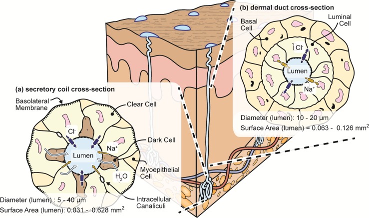
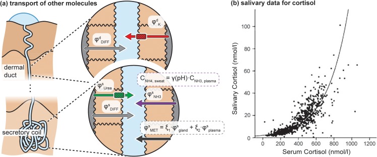
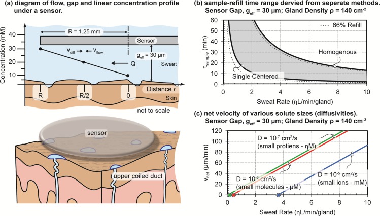
Non-invasive and accurate access of biomarkers remains a holy grail of the biomedical community. Human eccrine sweat is a surprisingly biomarker-rich fluid which is gaining increasing attention. This is especially true in applications of continuous bio-monitoring where other biofluids prove more challenging, if not impossible. However, much confusion on the topic exists as the microfluidics of the eccrine sweat gland has never been comprehensively presented and models of biomarker partitioning into sweat are either underdeveloped and/or highly scattered across literature. Reported here are microfluidic models for eccrine sweat generation and flow which are coupled with review of blood-to-sweat biomarker partition pathways, therefore providing insights such as how biomarker concentration changes with sweat flow rate. Additionally, it is shown that both flow rate and biomarker diffusion determine the effective sampling rate of biomarkers at the skin surface (chronological resolution). The discussion covers a broad class of biomarkers including ions (Na(+), Cl(-), K(+), NH4 (+)), small molecules (ethanol, cortisol, urea, and lactate), and even peptides or small proteins (neuropeptides and cytokines). The models are not meant to be exhaustive for all biomarkers, yet collectively serve as a foundational guide for further development of sweat-based diagnostics and for those beginning exploration of new biomarker opportunities in sweat.


Figures

FIG. 1.
FIG. 1.
Structure of the human sweat gland showing the (a) skin cross-section and (b) microfluidic equivalent.
FIG. 2.
FIG. 2.
Cross-sectional and top-down illustration of the (a) secretory coil and (b) dermal duct.
FIG. 3.
FIG. 3.
Images of the finger tip of author Swaile showing individual gland firing, pulsation, and sweat bead recession. The dotted line is the perimeter of the generated sweat.
FIG. 4.
FIG. 4.
Transport model for two common electrolytes. (a) Illustrative transport model for sodium, chloride, and water in the eccrine gland. (b) Biomarker surface concentration dependence upon sweat rate, in part adapted from Ref. . It is speculated that increased solute concentrations at very low sweat rates are due to evaporation and fluid absorption on the skin.
FIG. 5.
FIG. 5.
Transport of molecules other than sodium or chloride. (a) Illustrative partitioning model for potassium, ammonia, urea, molecules of metabolic origin, and a placeholder for other diffusively partitioning biomarkers. (b) Relationship between salivary and serum cortisol. Reprinted with permission from Hellhammer et al., “Salivary cortisol as a biomarker in stress research,” Psychoneuroendocrinology 34, 163–171 (2009). Copyright 2009 Elsevier.
FIG. 6.
FIG. 6.
Estimation of the minimum sampling time (refresh rate) for a sweat biosensor. (a) Representation of the case where there is a linear concentration gradient below the sensor with flow (vflow) and diffusional (vdiff) velocities in opposition. (b) The effective refill time from two separate derivation methods, see Appendix B in the supplementary material. (c) Simple, net molecular velocity underneath the sensor. Please see Appendix B for a full derivation.

References

    1. Sato K. Reviews of Physiology, Biochemistry and Pharmacology ( Springer, 1977), Vol. 79, pp. 51–131. - PubMed
    1. Sato K., Kang W. H., Saga K., and Sato K. T., “ Biology of sweat glands and their disorders. I. Normal sweat gland function,” J. Am. Acad. Dermatol. 20, 537–563 (1989). 10.1016/S0190-9622(89)70063-3 - DOI - PubMed
    1. De Giovanni N. and Fucci N., “ The current status of sweat testing for drugs of abuse: A review,” Curr. Med. Chem. 20, 545–561 (2013). 10.2174/092986713804910139 - DOI - PubMed
    1. Kidwell D. A., Holland J. C., and Athanaselis S., “ Testing for drugs of abuse in saliva and sweat,” J. Chromatogr. B 713, 111–135 (1998). 10.1016/S0378-4347(97)00572-0 - DOI - PubMed
    1. Mena-Bravo A. and Luque de Castro M. D., “ Sweat: A sample with limited present applications and promising future in metabolomics,” J. Pharm. Biomed. Anal. 90, 139–147 (2014). 10.1016/j.jpba.2013.10.048 - DOI - PubMed