Review
. 2015 Feb 1;128(3):431-9.
doi: 10.1242/jcs.159400.

Connections matter--how viruses use cell–cell adhesion components

Review

Connections matter--how viruses use cell–cell adhesion components

Mathieu Mateo et al. J Cell Sci. .

Abstract

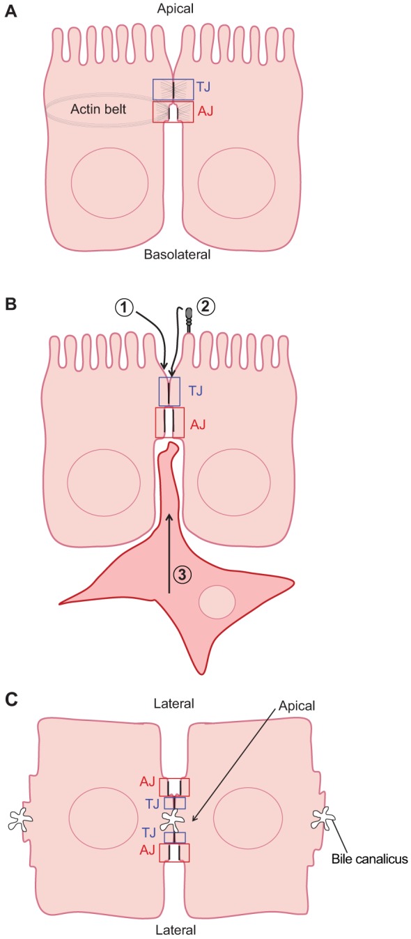
The epithelium is a highly organized type of animal tissue. Except for blood and lymph vessels, epithelial cells cover the body, line its cavities in single or stratified layers and support exchange between compartments. In addition, epithelia offer to the body a barrier to pathogen invasion. To transit through or to replicate in epithelia, viruses have to face several obstacles, starting from cilia and glycocalyx where they can be neutralized by secreted immunoglobulins. Tight junctions and adherens junctions also prevent viruses to cross the epithelial barrier. However, viruses have developed multiple strategies to blaze their path through the epithelium by utilizing components of cell–cell adhesion structures as receptors. In this Commentary, we discuss how viruses take advantage of the apical junction complex to spread. Whereas some viruses quickly disrupt epithelium integrity, others carefully preserve it and use cell adhesion proteins and their cytoskeletal connections to rapidly spread laterally. This is exemplified by the hidden transmission of enveloped viruses that use nectins as receptors. Finally, several viruses that replicate preferentially in cancer cells are currently used as experimental cancer therapeutics. Remarkably, these viruses use cell adhesion molecules as receptors, probably because--to reach tumors and metastases--ncolytic viruses must efficiently traverse or break epithelia.


Figures

Fig. 1.
Fig. 1.
Structure of different epithelial cells, and distinct modes of virus entry. (A) Representation of two polarized columnar epithelial cells making contact through the tight junction (TJ, blue box) and the adherens junction (AJ, red box). Indicated are the apical and basolateral sides as well as the actin belt, which is anchored to the AJ. (B) Three different strategies adopted by viruses to enter epithelial cells. Strategies 1 and 2 are for apical entry of viral particles. In strategy 1, the virus contacts a receptor within the TJ directly, whereas in strategy 2 a co-receptor transports the virus to the TJ. Strategy 3 allows basolateral entry of cell-associated viruses. The infected cell is shown with a thick red membrane, to imply the presence of the receptor-binding viral glycoproteins. Different viruses use one of these strategies (see text for details). (C) Schematic representation of two hepatocytes making contacts. Here, the apical surface corresponds to the bile canaliculus between the two cells. Two TJs and external AJs surround the canaliculus. The sinusoidal vessels in both orientations are lateral surfaces.
Fig. 2.
Fig. 2.
Main components of the tight and adherens junctions in polarized epithelial cells. (A) Tight junction with four transmembrane proteins (top to bottom): coxsackievirus and adenovirus receptor (CAR), junctional adhesion molecule (JAM), claudin and occludin. All these proteins are associated with the zonula occludens proteins ZO-1, ZO-2 and ZO-3 (yellow) through their cytosolic tails. ZO proteins link the transmembrane proteins to the actin cytoskeleton. (B) Adherens junction with two trasmembrane proteins, cadherins and nectins. Cadherins are linked to the F-actin cytoskeleton through α- and β-catenins (α and β). Nectins are linked to F-actin through the large protein afadin (AF6). AF6 and the catenins also interact.
Fig. 3.
Fig. 3.
Viruses use junctional proteins as receptors. (A) Viruses interacting with TJ proteins. Top to bottom: adenovirus and coxsackievirus interact with CAR, reovirus with JAM, and hepatitis C virus (HCV) interacts with both claudin and occludin. (B) Viruses interacting with AJ proteins. No virus interacts with cadherins. Herpes simplex virus (HSV) and other animal viruses of the same family interact with nectin-1 and nectin-2. Measles virus (MeV) and related animal morbilliviruses interact with nectin-4. Poliovirus interacts with Necl5.
Fig. 4.
Fig. 4.
Viral spread with or without particle formation. Three different strategies for virus spread are illustrated. In strategy 1, virus particles bud from the apical surface. Depending on their receptor location, the released particles may or not be able to re-enter the epithelium. Strategy 2 is similar to strategy 1, but viral particles are released into the intercellular space near the junctional complex where they are able to contact their receptor. In strategy 3, the virus does not form extracellular particles. This hypothetical strategy applies to enveloped viruses with glycoproteins that can fuse membranes, forming pores allowing the intercellular transfer of viral genomes. The infected cell is shown with a thick red membrane, to imply the presence of the receptor-binding viral glycoproteins. In strategy 2, measles virus and HSV nucleocapsids are enveloped (thick red membrane), whereas in strategy 3 they are not. RNA genomes are shown in red, DNA genomes in blue. Single stranded genomes are indicated by one wavy line, double stranded genomes by two wavy lines. Hexagons represent icosahedral capsids, circles represent membranes, helical capsids are not indicated. The three viruses in for strategy 1 are coxsackie, reo- and adenoviruses. The two viruses for strategies 2 and 3 are HSV and the measles virus (see text for further details).

References

    1. Andrus L., Marukian S., Jones C. T., Catanese M. T., Sheahan T. P., Schoggins J. W., Barry W. T., Dustin L. B., Trehan K., Ploss A. et al. (2011). Expression of paramyxovirus V proteins promotes replication and spread of hepatitis C virus in cultures of primary human fetal liver cells. Hepatology 54, 1901–1912 10.1002/hep.24557 - DOI - PMC - PubMed
    1. Antar A. A., Konopka J. L., Campbell J. A., Henry R. A., Perdigoto A. L., Carter B. D., Pozzi A., Abel T. W., Dermody T. S. (2009). Junctional adhesion molecule-A is required for hematogenous dissemination of reovirus. Cell Host Microbe 5, 59–71 10.1016/j.chom.2008.12.001 - DOI - PMC - PubMed
    1. Arora P., Kim E. O., Jung J. K., Jang K. L. (2008). Hepatitis C virus core protein downregulates E-cadherin expression via activation of DNA methyltransferase 1 and 3b. Cancer Lett. 261, 244–252 10.1016/j.canlet.2007.11.033 - DOI - PubMed
    1. Barclay A. N. (1999). Ig-like domains: evolution from simple interaction molecules to sophisticated antigen recognition. Proc. Natl. Acad. Sci. USA 96, 14672–14674 10.1073/pnas.96.26.14672 - DOI - PMC - PubMed
    1. Barton E. S., Forrest J. C., Connolly J. L., Chappell J. D., Liu Y., Schnell F. J., Nusrat A., Parkos C. A., Dermody T. S. (2001). Junction adhesion molecule is a receptor for reovirus. Cell 104, 441–451 10.1016/S0092-8674(01)00231-8 - DOI - PubMed

Publication types

LinkOut - more resources