. 2015 Aug 28;6(25):21395-405.
doi: 10.18632/oncotarget.4095.

BEX1 acts as a tumor suppressor in acute myeloid leukemia

Affiliations

BEX1 acts as a tumor suppressor in acute myeloid leukemia

Oscar Lindblad et al. Oncotarget. .

Abstract

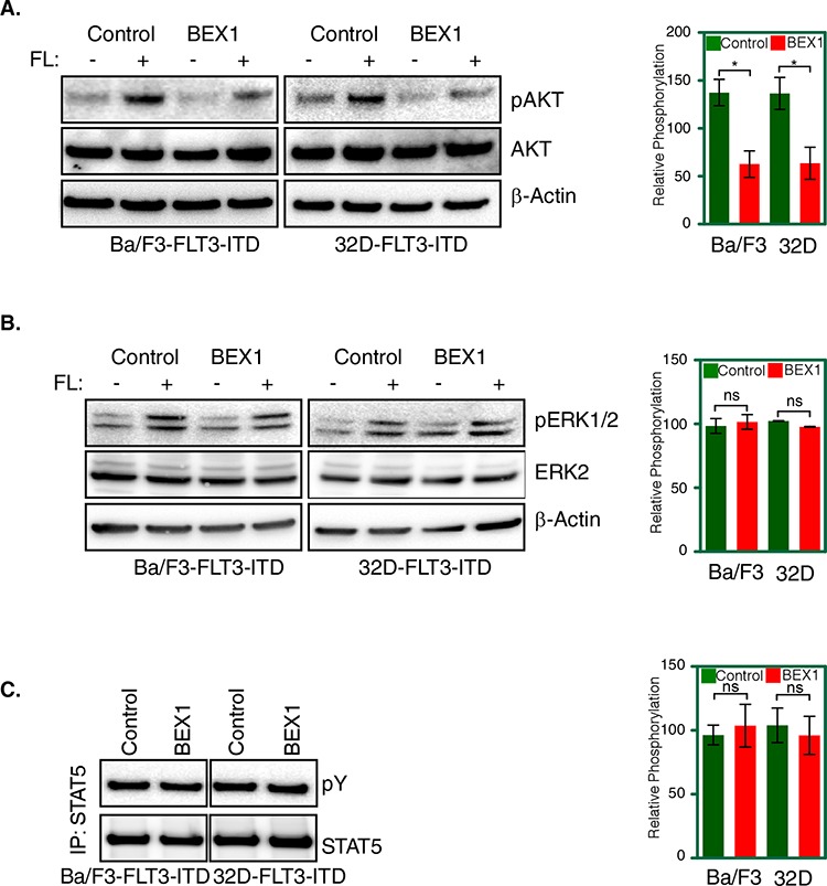
Acute myeloid leukemia (AML) is a heterogeneous disease of the myeloid lineage. About 35% of AML patients carry an oncogenic FLT3 mutant making FLT3 an attractive target for treatment of AML. Major problems in the development of FLT3 inhibitors include lack of specificity, poor response and development of a resistant phenotype upon treatment. Further understanding of FLT3 signaling and discovery of novel regulators will therefore help to determine additional pharmacological targets in FLT3-driven AML. In this report, we identified BEX1 as a novel regulator of oncogenic FLT3-ITD-driven AML. We showed that BEX1 expression was down-regulated in a group of AML patients carrying FLT3-ITD. Loss of BEX1 expression resulted in poor overall survival (hazard ratio, HR = 2.242, p = 0.0011). Overexpression of BEX1 in mouse pro-B and myeloid cells resulted in decreased FLT3-ITD-dependent cell proliferation, colony and tumor formation, and in increased apoptosis in vitro and in vivo. BEX1 localized to the cytosolic compartment of cells and significantly decreased FLT3-ITD-induced AKT phosphorylation without affecting ERK1/2 or STAT5 phosphorylation. Our data suggest that the loss of BEX1 expression in FLT3-ITD driven AML potentiates oncogenic signaling and leads to decreased overall survival of the patients.

Keywords: AKT; AML; FLT3; FLT3-ITD; apoptosis.


Conflict of interest statement

CONFLICTS OF INTEREST

The authors declare no conflict of interests.

Figures

Figure 1
Figure 1. Deregulated gene expression in MV4-11 and MOLM-13 cell lines
A. Deregulated gene expression patterns in MV4-11 and MOLM-13 cell lines. B. Up-regulated and down-regulated genes in MV4-11 versus MOLM-13 cell lines. C. Relative BEX1 expression in MOLM-13 and MV4-11 cell lines. D. The BEX1 expression is deregulated in AML patients. Data set GSE14468 was used.
Figure 2
Figure 2. Overall survival of AML patients with higher and lower BEX1 expression
Data set GSE14468 was used in this analysis. Z-score was used to divide higher (n = 50) and lower (n = 50) BEX1 expressing patients. A–D. Overall survival of AML patients with FLT3-ITD positive and BEX1 higher or lower expression (A), FLT3-ITD negative and BEX1 higher or lower expression (B), FLT3-ITD positive plus BEX1 higher versus FLT3-ITD negative plus BEX1 lower expression (C) and, FLT3-ITD negative plus BEX1 higher versus FLT3-ITD positive plus BEX1 lower expression (D).
Figure 3
Figure 3. GSEA showed enrichment of oncogenic pathways in lower BEX1 expressing cells and patients
Data set GSE14468 was used in this analysis. Z-score was used to divide higher (n = 50) and lower (n = 50) BEX1 expressing patients. A. MV4-11 cells display enrichment of several oncogenic pathways in comparison to MOLM-13 cells. B. AML patients with lower BEX1 expression showed enrichment of several oncogenic pathways compared to patients with higher BEX1 expression.
Figure 4
Figure 4. BEX1 expression inhibited cell proliferation, colony formation and enhanced apoptosis
Cells were washed three times with RPMI-1640 to remove IL3. A. Expression of BEX1 and FLT3-ITD in stably transfected Ba/F3 and 32D cells was measured by western blotting analysis. B. FLT3-ITD dependent cell proliferation in presence and absence of BEX1 expression was measured after 24 and 48 hours using stably transfected Ba/F3 and 32D cells. C. Stably transfected Ba/F3 and 32D cells were used to determine colony formation potential in the semi-solid medium. D. Apoptosis induced by BEX1 expression was measured using Annexin-V and 7-AAD kit.
Figure 5
Figure 5. BEX1 delayed tumor growth in mouse xenograft
Cells were washed three times with RPMI-1640 to remove IL3. A–D. Cells expressing BEX1 or control were xenografted into mice and tumor growth was monitored for 24 days. Tumor volume (A) and weight (B) in Ba/F3 cells as well as in 32D cells (tumor volume (C) and weight (D)) were analyzed.
Figure 6
Figure 6. BEX1 localized to the cytosol and did not affect FLT3-ITD stability
A. Localization of BEX1 was visualized by confocal microscope. FLT3-ITD was stained with PE-conjugated antibody and BEX1-FLAG was stained with Alexa Flour 647-conjugated anti-FLAG antibody. B. Cells were washed three times with RPMI-1640 to remove IL3. FLT3 degradation was measured in transfected Ba/F3 and 32D cells after 30 minutes of ligand stimulation.
Figure 7
Figure 7. BEX1 expression decreased AKT phosphorylation
Cells were washed three times with RPMI-1640 to remove IL3. A–B. Transfected Ba/F3 and 32D cells were stimulated with a ligand for 5 minutes before lysis. Total cell lysates were used for SDS-PAGE and western blotting analysis with AKT (A) and ERK (B) antibodies. Ligand stimulated samples were used for quantification. C. Cell lysates for stimulated and unstimulated cells were immunoprecipitated with an anti-STAT5 antibody followed by western blotting analysis.

References

    1. Gilliland DG, Griffin JD. The roles of FLT3 in hematopoiesis and leukemia. Blood. 2002;100:1532–1542. - PubMed
    1. Kabir NN, Rönnstrand L, Kazi JU. FLT3 mutations in patients with childhood acute lymphoblastic leukemia (ALL) Med Oncol. 2013;30:462. - PubMed
    1. Schnittger S, Schoch C, Dugas M, Kern W, Staib P, Wuchter C, Löffler H, Sauerland CM, Serve H, Büchner T, Haferlach T, Hiddemann W. Analysis of FLT3 length mutations in 1003 patients with acute myeloid leukemia: correlation to cytogenetics, FAB subtype, and prognosis in the AMLCG study and usefulness as a marker for the detection of minimal residual disease. Blood. 2002;100:59–66. - PubMed
    1. Fröhling S, Schlenk RF, Breitruck J, Benner A, Kreitmeier S, Tobis K, Döhner H, Döhner K leukemia AMLSGUAm. Prognostic significance of activating FLT3 mutations in younger adults (16 to 60 years) with acute myeloid leukemia and normal cytogenetics: a study of the AML Study Group Ulm. Blood. 2002;100:4372–4380. - PubMed
    1. Kiyoi H, Yanada M, Ozekia K. Clinical significance of FLT3 in leukemia. International journal of hematology. 2005;82:85–92. - PubMed

Publication types

MeSH terms