. 2015 Jun 9;65(22):2404-15.
doi: 10.1016/j.jacc.2015.03.573.

Differential Inhibition of Human Atherosclerotic Plaque-Induced Platelet Activation by Dimeric GPVI-Fc and Anti-GPVI Antibodies: Functional and Imaging Studies

Affiliations

Differential Inhibition of Human Atherosclerotic Plaque-Induced Platelet Activation by Dimeric GPVI-Fc and Anti-GPVI Antibodies: Functional and Imaging Studies

Janina Jamasbi et al. J Am Coll Cardiol. .

Abstract

Background: Glycoprotein VI (GPVI) is the essential platelet collagen receptor in atherothrombosis, but its inhibition causes only a mild bleeding tendency. Thus, targeting this receptor has selective antithrombotic potential.

Objectives: This study sought to compare compounds interfering with platelet GPVI-atherosclerotic plaque interaction to improve current antiatherothrombotic therapy.

Methods: Human atherosclerotic plaque-induced platelet aggregation was measured in anticoagulated blood under static and arterial flow conditions (550/s, 1,100/s, and 1,500/s). Inhibition by dimeric GPVI fragment crystallizable region of IgG (Fc) masking GPVI binding sites on collagen was compared with that of 3 anti-GPVI antibodies: BLO8-1, a human domain antibody; 5C4, a fragment antigen-binding (Fab fragment) of monoclonal rat immunoglobulin G; and m-Fab-F, a human recombinant sFab against GPVI dimers.

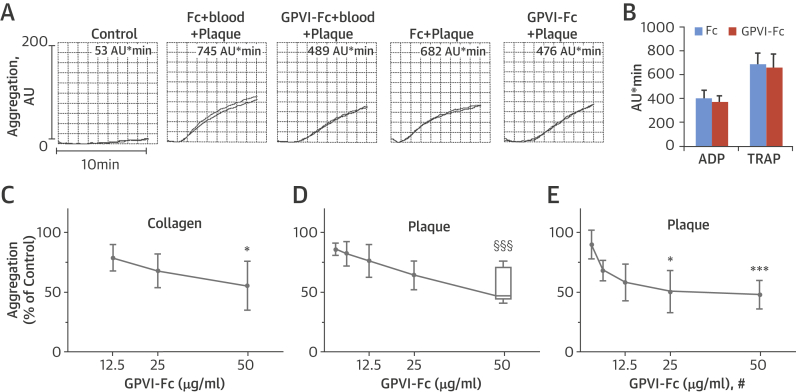
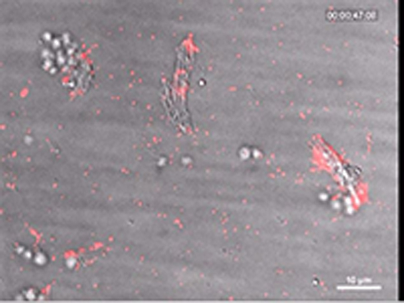
Results: GPVI-Fc reduced plaque-triggered platelet aggregation in static blood by 51%, BLO8-1 by 88%, and 5C4 by 93%. Under arterial flow conditions, BLO8-1 and 5C4 almost completely inhibited platelet aggregation while preserving platelet adhesion on plaque. Inhibition by GPVI-Fc, even at high concentrations, was less marked but increased with shear rate. Advanced optical imaging revealed rapid persistent GPVI-Fc binding to collagen under low and high shear flow, upstream and downstream of plaque fragments. At low shear particularly, platelets adhered in plaque flow niches to GPVI-Fc-free segments of collagen fibers and recruited other platelets onto aggregates via ADP and TxA2 release.

Conclusions: Anti-GPVI antibodies inhibit atherosclerotic plaque-induced platelet aggregation under static and flow conditions more effectively than GPVI-Fc. However, potent platelet inhibition by GPVI-Fc at a higher shear rate (1,500/s) suggests localized antithrombotic efficacy at denuded or fissured stenotic high-risk lesions without systemic bleeding. The compound-specific differences have relevance for clinical trials targeting GPVI-collagen interaction combined with established antiplatelet therapies in patients with spontaneous plaque rupture or intervention-associated plaque injury.

Keywords: antithrombotic; atherothrombosis; glycoprotein VI; plaque rupture.


Figures

Figure 1
Figure 1
Static Platelet Aggregation Attenuated by GPVI-Fc (A) Representative multiple electrode aggregometry tracings show plaque-induced platelet aggregation in blood pre-incubated with solvent, equimolar concentrations of Fc (16 μg/ml), and GPVI-Fc (50 μg/ml) (tracings 1 to 3). Plaque samples pre-incubated with 35-fold higher equimolar concentrations of Fc (560 μg/ml) or GPVI-Fc (1,750 μg/ml) for 3 min were added to blood yielding the same final concentrations (tracings 4 and 5). Numbers show cumulative aggregation (AU*min) measured from 0 to 10 min. (B) GPVI-Fc does not affect platelet aggregation stimulated by ADP (5 μM) or TRAP (15 μM) (mean ± SD, n = 4). Blood was pre-incubated with GPVI-Fc or Fc control protein in increasing concentrations before stimulation with collagen (0.5 μg/ml) (C) or with plaque (833 μg/ml) (D) for 5 min. (E) Plaque was pre-incubated with equimolar concentrations of GPVI-Fc (109, 219, 437, 875, and 1,750 μg/ml) or Fc for 3 min before added to blood, yielding the same final concentrations (#) as in D. *p < 0.05; ***p < 0.001 for GPVI-Fc versus control by 2-tailed paired Student t test, or §§§p < 0.001 by the Mann–Whitney U test. ADP = adenosine diphosphate; Fc = fragment crystallizable region; GPVI = glycoprotein VI; TRAP = thrombin receptor-activating peptide.
Figure 2
Figure 2
Inhibition of Atherosclerotic Plaque-Induced Platelet Deposition by GPVI-Fc and Anti-GPVI Antibodies Representative micrographs display platelet coverage of plaque at different times after start of blood flow at 550/s (A) (Online Videos 1 and 2) or 1,100/s (B). Blood was pre-incubated with mepacrine for platelet visualization (without = control) or with GPVI-Fc (50 μg/ml) or anti-GPVI antibodies 5C4 (1.25 μg/ml) or BLO8-1 (20 μg/ml). Enlarged insets = high magnification images. (C) Effect of GPVI-Fc, BLO8-1, or 5C4 on the time course of platelet deposition onto plaque from flowing blood at 3 different arterial shear rates (Online Videos 1 and 2). BLO8-1 and 5C4 curves are shown at blown-up scale (right). Mean ± SD of 5 to 12 experiments. Secondary pair comparisons between treatments were significant for control versus Blo8-1 (**p < 0.01), 5C4 (**), and GPVI-Fc (*p < 0.05). Abbreviations as in Figure 1.
Figure 2
Figure 2
Inhibition of Atherosclerotic Plaque-Induced Platelet Deposition by GPVI-Fc and Anti-GPVI Antibodies Representative micrographs display platelet coverage of plaque at different times after start of blood flow at 550/s (A) (Online Videos 1 and 2) or 1,100/s (B). Blood was pre-incubated with mepacrine for platelet visualization (without = control) or with GPVI-Fc (50 μg/ml) or anti-GPVI antibodies 5C4 (1.25 μg/ml) or BLO8-1 (20 μg/ml). Enlarged insets = high magnification images. (C) Effect of GPVI-Fc, BLO8-1, or 5C4 on the time course of platelet deposition onto plaque from flowing blood at 3 different arterial shear rates (Online Videos 1 and 2). BLO8-1 and 5C4 curves are shown at blown-up scale (right). Mean ± SD of 5 to 12 experiments. Secondary pair comparisons between treatments were significant for control versus Blo8-1 (**p < 0.01), 5C4 (**), and GPVI-Fc (*p < 0.05). Abbreviations as in Figure 1.
None
Online Video 1
None
Online Video 2
None
Online Video 3
Figure 3
Figure 3
Dynamics of GPVI-Fc Binding, Platelet Adhesion, and Aggregate Formation Onto Atherosclerotic Plaque Material Under Flow (A) GPVI-Fc (50 μg/ml final concentration) labeled with phycoerythrin (PE)–conjugated anti-human Fc antibody (red) and added to blood before perfusion rapidly bound to plaque homogenate. Binding was similar at low and high shear rates (550/s and 1,500/s). Deposition of platelets (green; labeled with DiOC6) lagged behind at low shear and was inhibited at high shear. (B) GPVI-Fc-PE (red) binds up- and downstream of plaque fragments (gray) and to small plaque pieces not detectable by differential interference contrast (DIC) (Online Videos 4, 5, and 7). Platelet adhesion and aggregate formation (gray) is observed only downstream. Phase contrast (DIC) images of platelet (gray) and GPVI-Fc-PE binding (red) to plaque at different times after start of blood perfusion at low shear rate (550/s). Rows 1 and 2: A single platelet (upper arrow) rolls over PE-labeled GPVI-Fc (lower arrow) bound to a piece of plaque material. Rows 3 and 4: Platelet aggregate formation starting from a single adhering platelet (arrow) in a flow niche downstream of plaque. Rows 1 and 3: Overlay of DIC (plaque/platelets) and fluorescence (PE-labeled GPVI-Fc) images; rows 2 and 4: fluorescence images of PE-labeled GPVI-Fc. Bar = 5 μm. Abbreviations as in Figure 1.
None
Online Video 4
None
Online Video 5
None
Online Video 6
None
Online Video 7
Figure 4
Figure 4
Platelets Adhere Downstream to Sites of Plaque Collagen Not Occupied by But in Close Proximity to GPVI-Fc as Revealed by SIM Imaging Plaque homogenates pre-stained with anti-collagen types I and III antibody (Ab) and Alexa Fluor 405 conjugated second Ab were perfused with blood containing Alexa Fluor 594–labeled GPVI-Fc (red) (50 μg/ml) and abciximab (to block platelet aggregation) at a shear rate of 550/s. After 3 min of flow, samples were fixed, and platelets (green) were stained with anti-CD41 Ab and DyLight 488 conjugated second Ab. Structured illumination microscopy fluorescence micrographs were taken of the subsequent 0.2-μm sections of the sample, and 3-dimensional reconstructions were made with ImagePro Premier 3D (version 9.1, Media Cybernetics, Rockville, Maryland). (Top) Three-dimensional overview of the sample (thickness, 3.6 μm). (Bottom) Magnified subvolumes of the sample at 2 z positions from bottom to top (z1 = 1.0 to 1.6 μm; z2 = 1.6 to 2.2 μm) revealing platelet adhesion to discrete sites of plaque collagen (blue, arrows). Black arrow shows direction of blood flow. Image is representative of 7 others (see also Online Video 8). SIM = structured illumination microscopy.
None
Online Video 8
Figure 5
Figure 5
Residual Platelet Aggregate Formation in the Presence of GPVI-Fc Is Inhibited by Blockade of Platelet Cyclooxygenase and the P2Y12 Receptor Blood was pre-incubated for 5 min with buffer (control), GPVI-Fc (50 μg/ml), the P2Y12 receptor antagonist AR-C69931 (1 μM) added to blood containing acetylsalicylic acid (ASA) (1 mM), alone or in combination with GPVI-Fc. Blood was perfused over plaque at a shear rate of 550/s. Mean ± SD (n = 5). p < 0.005 for treatment with ASA + P2Y12 antagonist + GPVI-Fc (endpoint) compared with ASA + P2Y12 antagonist and GPVI-Fc. Abbreviations as in Figure 1.
None
Online Video 9
Central Illustration
Differential Inhibition of Plaque-Induced Platelet Aggregate Formation by GPVI-Fc and Anti-GPVI Antibodies Glycoprotein VI is an essential platelet collagen receptor. (A) Dimeric fusion protein GPVI-Fc binds to exposed GPO sites of collagen up- and downstream after plaque rupture and competes with platelet GPVI dimer. Only a few GPO sites unoccupied by GPVI-Fc are needed to induce efficient platelet GPVI signaling with subsequent ADP and TxA2 release (broken white curved arrow) mediating stable platelet adhesion and aggregation in flow niches under low shear (top), but not under high shear at sites of high-risk stenotic lesion ruptures as ADP and TxA2 are flushed away (broken white arrow) due to the high-flow velocity (bottom); white arrow = direction of blood flow. (B) Anti-GPVI antibodies bind to platelets in the circulation and inhibit platelet aggregation after plaque rupture independently of flow and stenosis. ADP = adenosine diphosphate; Fc = fragment crystallizable region of IgG; GPO = glycine-proline-hydroxyproline; GPVI = glycoprotein VI; GPVIex = external domain of GPVI; Plt = platelet; TxA2 = thromboxane A2.

References

    1. Fuster V., Moreno P.R., Fayad Z.A., Corti R., Badimon J.J. Atherothrombosis and high-risk plaque: part I: evolving concepts. J Am Coll Cardiol. 2005;46:937–954. - PubMed
    1. Badimon L., Vilahur G. Thrombosis formation on atherosclerotic lesions and plaque rupture. J Int Med. 2014;276:618–632. - PubMed
    1. Reininger A.J., Bernlochner I., Penz S.M., et al. A 2-step mechanism of arterial thrombus formation induced by human atherosclerotic plaques. J Am Coll Cardiol. 2010;55:1147–1158. - PubMed
    1. van Zanten G.H., de Graaf S., Slootweg P.J., et al. Increased platelet deposition on atherosclerotic coronary arteries. J Clin Invest. 1994;93:615–632. - PMC - PubMed
    1. Penz S., Reininger A.J., Brandl R., et al. Human atheromatous plaques stimulate thrombus formation by activating platelet glycoprotein VI. FASEB J. 2005;19:898–909. - PubMed

MeSH terms

Substances