. 2015 Jun 4;16(6):627-38.
doi: 10.1016/j.stem.2015.04.013.

Krt19(+)/Lgr5(-) Cells Are Radioresistant Cancer-Initiating Stem Cells in the Colon and Intestine

Affiliations

Krt19(+)/Lgr5(-) Cells Are Radioresistant Cancer-Initiating Stem Cells in the Colon and Intestine

Samuel Asfaha et al. Cell Stem Cell. .

Abstract

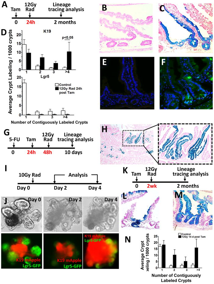
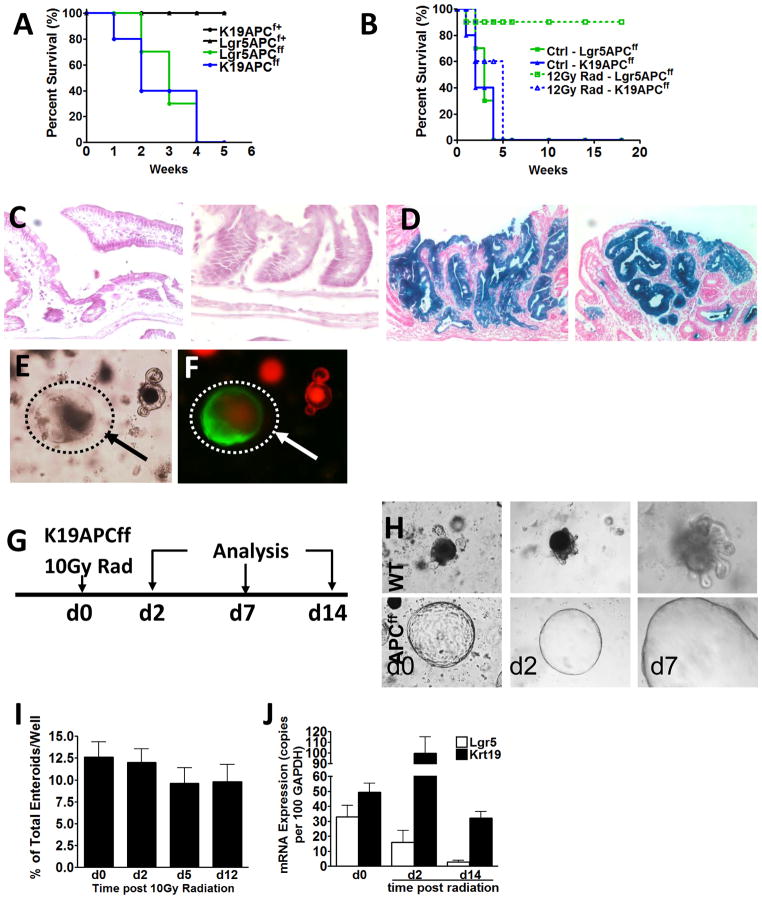
Epithelium of the colon and intestine are renewed every 3 days. In the intestine there are at least two principal stem cell pools. The first contains rapid cycling crypt-based columnar (CBC) Lgr5(+) cells, and the second is composed of slower cycling Bmi1-expressing cells at the +4 position above the crypt base. In the colon, however, the identification of Lgr5(-) stem cell pools has proven more challenging. Here, we demonstrate that the intermediate filament keratin-19 (Krt19) marks long-lived, radiation-resistant cells above the crypt base that generate Lgr5(+) CBCs in the colon and intestine. In colorectal cancer models, Krt19(+) cancer-initiating cells are also radioresistant, while Lgr5(+) stem cells are radiosensitive. Moreover, Lgr5(+) stem cells are dispensable in both the normal and neoplastic colonic epithelium, as ablation of Lgr5(+) stem cells results in their regeneration from Krt19-expressing cells. Thus, Krt19(+) stem cells are a discrete target relevant for cancer therapy.


Conflict of interest statement

The authors do not have any conflicts of interests to declare.

Figures

Figure 1
Figure 1. Krt19 mRNA localizes to the colonic and intestinal stem cell zone and marks long-lived stem cells
Krt19 mRNA is expressed in the isthmus extending down to the +4 position of the colonic (A) and (B) intestinal crypt. High magnification images of the crypt base are shown as insets. Krt19-mApple+ cells in the colon (C) and small intestine (D) of Krt19-mApple reporter mice show expression similar to in situ. 24 h post tamoxifen, β-gal+ colonic (E) and intestinal (F) crypts in Krt19-CreERT;R26RLacZ mice also show expression identical to in situ. Lineage tracing in the colon (G) of Krt19-CreERT;R26RLacZ mice. High magnification images of the crypt base are shown at 24 hours and 52 weeks following tamoxifen (6 mg p.o.). Low magnification images of lineage tracing in the colon (H) of Krt19-CreERT;R26RLacZ 26 weeks following tamoxifen (n ≥ 7 per group). Bright-field (6 and 24 hours after culture) (I) and 2-photon images (24 hours and 9 days following tamoxifen (6 mg p.o.)) (J) of colonic crypts from Krt19-CreERT; R26-mT/mG mice (n ≥ 4 per group) cultured in vitro. Lineage tracing in the intestine (K) in Krt19-CreERT/R26RLacZ mice with high magnification images of the crypt base shown at 24 hours to 52 weeks following tamoxifen (6 mg p.o.). Note black arrows that show Krt19-derived CBCs at 7 days and Paneth cells at 16 and 52 weeks. Low magnification images of lineage tracing in the intestine (L) of Krt19-CreERT/R26RLacZ 26 weeks following tamoxifen (n ≥ 7 per group). See also Figure S1 and S2.
Figure 2
Figure 2. Krt19+ cells are located above Lgr5+ crypt base columnar cells in the colon and intestine
Colocalization of Krt19 mRNA expressing cells (red) detected by in situ and Lgr5-GFP+ cells (green) in the colon (A) and small intestine (B) of Lgr5-EGFP-IRES-CreERT2 mice. White arrows denote double positive cells. (C) Average cell position of Krt19 mRNA expressing (red) and Lgr5-EGFP+ (green) cells within the colonic (top panel) and intestinal (bottom panel) crypt. Colocalization of Ki67 and Lgr5-EGFP+ cells (D) versus Ki67 and Krt19-EGFP+ cells 12 h after tamoxifen (E) in Lgr5-EGFP-IRES-CreERT2 or Krt19-CreERT;ROSA26-mG/mT mice, respectively. Yellow arrow shows a rare double positive (Ki67+, Lgr5-GFP+) cells and white arrow shows a rare Krt19+, Ki67 cells. Quantification of double positive Ki67+Krt19+ cells (red bars) versus Ki67+Lgr5-GFP+ cells (green bars) (F). Representative images of the colon (G) and SI (H) of Krt19-mApple;Lgr5-EGFP-IRES-CreERT2 dual reporter mice showing no overlap of Krt19-mApple+ and Lgr5-GFP+ cells in the colon and rare Krt19-mApple+/Lgr5-GFP+ double positive cells higher in the crypt of the SI. FACS plot (I) and quantification (J) of colonic and intestinal Krt19-mApple+ and Lgr5-GFP+ positive cells from Krt19-mApple;Lgr5-EGFP-IRES-CreERT2 mice. Schematic diagram of the intestinal crypt demonstrating the location of Krt19-expressing cells in relation to Lgr5+ crypt based columnar cells (K). mRNA expression levels of + 4 stem cell (BMi1, Hopx and Lrig1) and progenitor (Dll1) markers among Krt19-mApple+, Lgr5-GFP+ and Krt19-mApple+/Lgr5-GFP+ double positive cell populations (L). See also Figure S3.
Figure 3
Figure 3. Krt19+ stem cells render Lgr5+ stem cells dispensable in the colon and small intestine
Tamoxifen protocol (A) used to analyze Krt19+ and Lgr5+ cells in Lgr5-DTR-EGFP;Krt19-CreERT/R26RTomato mice 24h post tamoxifen. Images of the colon and SI 24h post tamoxifen are shown (B). Diphtheria toxin (DT) ablation regimen (C) used in Lgr5-DTR-EGFP;Krt19-CreERT/R26RTomato mice showing Krt19+ stem cells (red) render Lgr5+ cells (green) dispensable in the colon and SI (D). Quantification of Krt19+ stem cell lineage tracing efficiency in the presence or absence of Lgr5+ stem cells (E) is shown. DT induced Lgr5+ cells ablation and 5-FU induced transit amplifying (TA) cell ablation regimen (F) used in Lgr5-DTR-EGFP;Krt19-CreERT/R26RTomato mice. (G) Representative high power view of Ki67+ cells in the colon of control (left) versus 5-FU treated (right) mice. Quantification of Ki67+ cells in the colon or intestine of control versus 5-FU treated mice (H). Krt19+ stem cells (red) render Lgr5+ cells (green) dispensable in the colon and SI (J) in spite of 5-FU ablation of TA cells. Quantification of Krt19+ stem cell lineage tracing efficiency in the presence or absence of DT and/or 5-FU stem cells (I) is shown (n ≥ 6 per group). See also Figure S4 and S5.
Figure 4
Figure 4. Krt19+ cells expand in response to injury and display relative radioresistance compared to Lgr5+ stem cells
To examine the radiosensitivity of Krt19+ and Lgr5+ stem cells, mice were irradiated 24 h after tamoxifen and lineage tracing examined 2 months following tamoxifen (A). Representative β-gal+ intestinal crypts in Lgr5-EGFP-IRES-CreERT2;R26RLacZ (B) versus Krt19-CreERT;R26RLacZ (C) mice irradiated (12 Gy) 24 h post tamoxifen. Quantification of contiguously labeled β-gal+ Krt19 (top) versus Lgr5 (bottom) labeled crypts following irradiation 24h after tamoxifen (D). Representative small intestinal crypt-villus image of Lgr5-EGFP-IRES-CreERT2;R26RLacZ mice following high dose radiation exposure demonstrating the disappearance of Lgr5-EGFP+ crypt based columnar cells 24 h following irradiation (E) and re-emergence of EGFP+ CBCs 7 days following irradiation (F). In vivo 5-FU (150 mg/kg) protocol used to examine the effects of TA cell ablation on Krt19+ stem cell lineage tracing (G). Representative low (left) and high (right) power images of Krt19+ cell lineage tracing in Krt19-CreERT;R26RLacZ mice treated with 5-FU and examined 8 d post radiation (H). In vitro radiation protocol used to examine the effects of radiation injury on intestinal Krt19 and Lgr5 stem cell populations (I). Bright-field (top) and fluorescent (bottom) images of intestinal enteroids from Krt19-mApple+/Lgr5-GFP+ double transgenic mice cultured in vitro pre and post radiation (10Gy) (J). Radiation protocol used to examine Lgr5 derived lineage tracing in Lgr5-EGFP-IRES-CreERT2/R26RLacZ mice two weeks after tamoxifen (K). β-gal+ intestinal crypts from control (L) versus irradiated (M) Lgr5-EGFP-IRES-CreERT2;R26RLacZ mice. Quantification of contiguously labeled β-gal+ Lgr5 labeled crypts following irradiation 2 weeks after tamoxifen (N); (n ≥ 5 per group). See also Figure S6.
Figure 5
Figure 5. Radioresistant Krt19+ cancer initiating cells are functionally distinct from Lgr5+ stem cells
Krt19+ and Lgr5+ stem cells both serve as cancer initiating cells resulting in rapid mortality in Krt19-CreERT;R26-LacZ;ApcF/F or Lgr5-EGFP-IRES-CreERT2;R26-LacZ;ApcF/F mice (A). Lgr5-EGFP-IRES-CreERT2;R26-LacZ;ApcF/F mice irradiated 24h after tamoxifen show no mortality (B) and normal non-lineage traced intestine (left) and colon (right) (C). In contrast, Krt19-CreERT;R26-LacZ;ApcF/F mice irradiated 24h after tamoxifen continue to show rapid mortality (B) from intestinal (left) and colonic (right) tumors (D); (n ≥ 6 per group). Bright-field (E) and fluorescent (F) images of intestinal crypts from Krt19-CreERT;R26-mT/mG;ApcF/F cultured in vitro 24 h after tamoxifen. Note arrows point to recombined GFP+ Apc floxed Krt19+ cells appearing as spheroid structures. In vitro radiation protocol used to examine the effects of radiation injury on intestinal Krt19+ cell derived Apc floxed tumor populations (G). Krt19+ cell derived Apc floxed tumor populations pre (d0) and post (day 2 and day 7) radiation (10 Gy) (H). Quantification of surviving Krt19+ cell derived APC floxed enteroids pre and post radiation injury (I). Krt19 and Lgr5 mRNA expression levels in Krt19+ cell derived Apc floxed enteroids pre and post radiation injury (J).
Figure 6
Figure 6. Lgr5+ stem cells are radiosensitive within colonic tumors unlike Krt19+ cells that are radioresistant
In vivo Krt19 and Lgr5 mRNA in Apc floxed tumors pre or post (24h and 7 d) radiation (A); * indicates p<0.05 vs control; (n ≥ 4 per group). Lgr5-GFP+ (B) and Krt19 immunopositive (C) cells in Apc floxed tumors pre or post radiation induced targeting of Lgr5+ cells. In vitro Lgr5+ cell ablation protocol used to examine the dispensability of Lgr5+ cells in Krt19+ cell derived Apc floxed enteroids from Lgr5-EGFP-DTR;Krt19-CreERT;ApcF/F mice (D). Bright-field images of intestinal enteroids from Lgr5-EGFP-DTR;Krt19-CreERT;ApcF/F mice pre (day 0) and post (day 2 and day 7) DT ablation of Lgr5+ cells (E). Quantification of surviving Krt19+ cell derived WT and APC floxed enteroids pre and post Lgr5+ cell ablation (F). Krt19 and Lgr5 mRNA expression levels in Krt19+ cell derived APC floxed enteroids pre and post (48 h) Lgr5+ cell ablation (G); ** indicates p<0.01 vs control; (n ≥ 4 per group). Bright-field images of Krt19+ cell derived APC floxed intestinal enteroids from Lgr5-EGFP-DTR;Krt19-CreERT;ApcF/F mice cultured in the presence (−DT) or absence (+DT) of Lgr5+ cell ablation (H). Quantification of surviving Krt19+ cell derived APC floxed enteroids pre and post Lgr5+ cell ablation and cultured in the presence/absence of growth factors (I). Krt19 and Lgr5 mRNA expression levels in Krt19+ cell derived APC floxed enteroids grown in the presence or absence of standard growth factors (R-spondin and noggin), pre and post (48 h) Lgr5+ cell ablation (J); (n ≥ 4 per group).

References

    1. Barker N, Ridgway RA, van Es JH, van de Wetering M, Begthel H, van den Born M, Danenberg E, Clarke AR, Sansom OJ, Clevers H. Crypt stem cells as the cells-of-origin of intestinal cancer. Nature. 2009;457:608–611. - PubMed
    1. Barker N, van Es JH, Kuipers J, Kujala P, van den Born M, Cozijnsen M, Haegebarth A, Korving J, Begthel H, Peters PJ, et al. Identification of stem cells in small intestine and colon by marker gene Lgr5. Nature. 2007;449:1003–1007. - PubMed
    1. Brembeck FH, Moffett J, Wang TC, Rustgi AK. The keratin 19 promoter is potent for cell-specific targeting of genes in transgenic mice. Gastroenterology. 2001;120:1720–1728. - PubMed
    1. Doetsch F, Caille I, Lim DA, Garcia-Verdugo JM, Alvarez-Buylla A. Subventricular zone astrocytes are neural stem cells in the adult mammalian brain. Cell. 1999;97:703–716. - PubMed
    1. Fearon ER, Vogelstein B. A genetic model for colorectal tumorigenesis. Cell. 1990;61:759–767. - PubMed

Publication types