. 2015 Jun 5;10(6):e0129152.
doi: 10.1371/journal.pone.0129152. eCollection 2015.

Quantitative Analyses of Force-Induced Amyloid Formation in Candida albicans Als5p: Activation by Standard Laboratory Procedures

Affiliations

Quantitative Analyses of Force-Induced Amyloid Formation in Candida albicans Als5p: Activation by Standard Laboratory Procedures

Cho X J Chan et al. PLoS One. .

Abstract

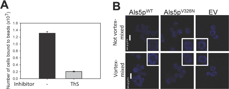
Candida albicans adhesins have amyloid-forming sequences. In Als5p, these amyloid sequences cluster cell surface adhesins to create high avidity surface adhesion nanodomains. Such nanodomains form after force is applied to the cell surface by atomic force microscopy or laminar flow. Here we report centrifuging and resuspending S. cerevisiae cells expressing Als5p led to 1.7-fold increase in initial rate of adhesion to ligand coated beads. Furthermore, mechanical stress from vortex-mixing of Als5p cells or C. albicans cells also induced additional formation of amyloid nanodomains and consequent activation of adhesion. Vortex-mixing for 60 seconds increased the initial rate of adhesion 1.6-fold. The effects of vortex-mixing were replicated in heat-killed cells as well. Activation was accompanied by increases in thioflavin T cell surface fluorescence measured by flow cytometry or by confocal microscopy. There was no adhesion activation in cells expressing amyloid-impaired Als5pV326N or in cells incubated with inhibitory concentrations of anti-amyloid dyes. Together these results demonstrated the activation of cell surface amyloid nanodomains in yeast expressing Als adhesins, and further delineate the forces that can activate adhesion in vivo. Consequently there is quantitative support for the hypothesis that amyloid forming adhesins act as both force sensors and effectors.


Conflict of interest statement

Competing Interests: The authors have declared that no competing interests exist.

Figures

Fig 1
Fig 1. The effects of vortex-mixing on adhesion and aggregation of S. cerevisiae.
Cells carrying an empty vector or expressing Als5pWT or Als5pV326N were vortex-mixed or not, then aggregated for 10 minutes with heat-denatured BSA-coated magnetic beads: (A) Bright-field micrographs of the cells. The dark-colored beads are 1 μm in diameter. Scale bars represent 20μm. (B) Quantification of cells adhering to beads. Error bars represent s.d. for n = 4. A student t-test was performed: *** represents p<0.001. (C) Time course for activation of Als5p-expressing cells by vortex mixing at 2500 rpm. Error bars represent s.e.m. for n = 8.
Fig 2
Fig 2. (A) Effects of anti-amyloid dye on vortex-activated adhesion and aggregation of S. cerevisiae cells expressing Als5pWT.
A 10- minute aggregation assay in the absence or presence of 0.2 mM ThS. (B) Effects of vortex mixing on surface amyloid nanodomains. Confocal micrographs of cells stained with 500 nM ThT without vortex mixing (top row) or after 5 min vortex mixing (bottom row). Scale bars represent 5μm.
Fig 3
Fig 3. Effect of vortex-mixing on aggregation and surface nanodomains on C. albicans SC5314 cells.
(A) Effects of vortex mixing on initial aggregation. Scale bars represent 100μm. (B) Number of cells bound to beads after vortex mixing for different times. Error bars represent standard deviation for n = 3. (C) ThT staining of control and vortex-mixed SC5314 cells. Scale bars represent 5μm. (D) Inhibition of aggregation with amyloid dyes 0.2mM ThS and 0.5mM Congo red after vortex-mixing.
Fig 4
Fig 4. Effect of centrifugation on aggregation.
Cells were grown in buffered medium, and aggregated in 10 min. assays. Some of the cells were centrifuged (3200 x g, 3mins) and resuspended before assay, and some of the centrifuged cells were also vortex mixed (2500 rpm, 1 min.) before assay.

References

    1. Otoo HN, Lee KG, Qiu W, Lipke PN. Candida albicans Als adhesins have conserved amyloid-forming sequences. Eukaryot Cell. 2008;7: 776–782. 10.1128/EC.00309-07 - DOI - PMC - PubMed
    1. Ramsook CB, Tan C, Garcia MC, Fung R, Soybelman G, Henry R, et al. Yeast cell adhesion molecules have functional amyloid-forming sequences. Eukaryot Cell. 2010;9: 393–404. 10.1128/EC.00068-09; 10.1128/EC.00068-09. - DOI - PMC - PubMed
    1. Fernandez-Escamilla AM, Rousseau F, Schymkowitz J, Serrano L. Prediction of sequence-dependent and mutational effects on the aggregation of peptides and proteins. Nat Biotechnol. 2004;22: 1302–1306. 10.1038/nbt1012 - DOI - PubMed
    1. Garcia MC, Lee JT, Ramsook CB, Alsteens D, Dufrene YF, Lipke PN. A role for amyloid in cell aggregation and biofilm formation. PLoS One. 2011;6: e17632 10.1371/journal.pone.0017632; 10.1371/journal.pone.0017632. - DOI - PMC - PubMed
    1. Alsteens D, Garcia MC, Lipke PN, Dufrene YF. Force-induced formation and propagation of adhesion nanodomains in living fungal cells. Proc Natl Acad Sci U S A. 2010;107: 20744–20749. 10.1073/pnas.1013893107; 10.1073/pnas.1013893107. - DOI - PMC - PubMed

Publication types

LinkOut - more resources