. 2015 Jun 5;10(6):e0128359.
doi: 10.1371/journal.pone.0128359. eCollection 2015.

Hyperglycemia and Diabetes Downregulate the Functional Expression of TRPV4 Channels in Retinal Microvascular Endothelium

Affiliations

Hyperglycemia and Diabetes Downregulate the Functional Expression of TRPV4 Channels in Retinal Microvascular Endothelium

Kevin Monaghan et al. PLoS One. .

Abstract

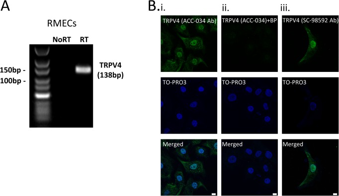
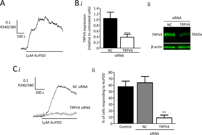
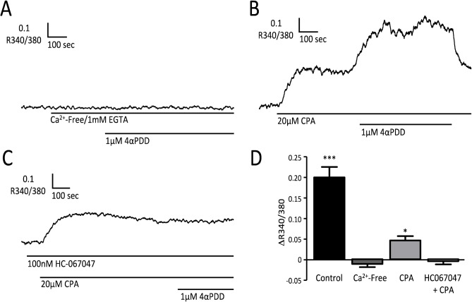
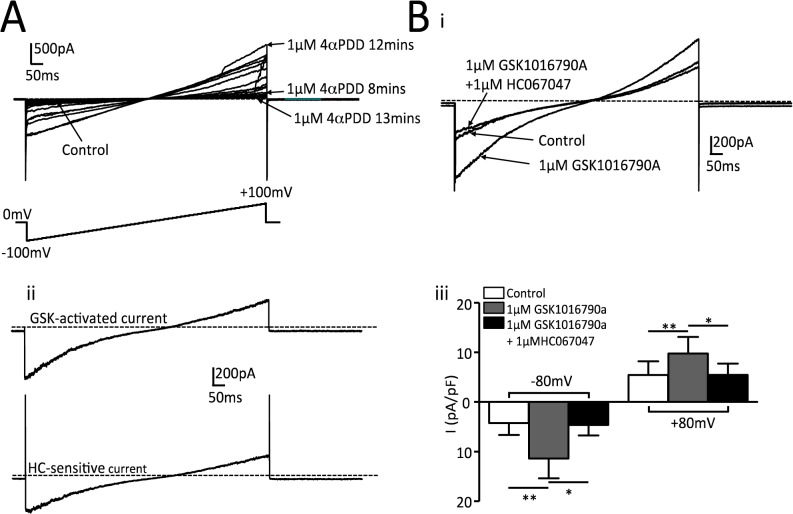
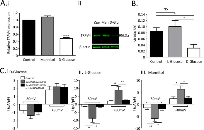
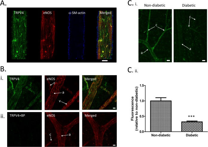
Retinal endothelial cell dysfunction is believed to play a key role in the etiology and pathogenesis of diabetic retinopathy. Numerous studies have shown that TRPV4 channels are critically involved in maintaining normal endothelial cell function. In the current paper, we demonstrate that TRPV4 is functionally expressed in the endothelium of the retinal microcirculation and that both channel expression and activity is downregulated by hyperglycaemia. Quantitative PCR and immunostaining demonstrated molecular expression of TRPV4 in cultured bovine retinal microvascular endothelial cells (RMECs). Functional TRPV4 activity was assessed in cultured RMECs from endothelial Ca2+-responses recorded using fura-2 microfluorimetry and electrophysiological recordings of membrane currents. The TRPV4 agonist 4α-phorbol 12,13-didecanoate (4-αPDD) increased [Ca2+]i in RMECs and this response was largely abolished using siRNA targeted against TRPV4. These Ca2+-signals were completely inhibited by removal of extracellular Ca2+, confirming their dependence on influx of extracellular Ca2+. The 4-αPDD Ca2+-response recorded in the presence of cyclopiazonic acid (CPA), which depletes the intracellular stores preventing any signal amplification through store release, was used as a measure of Ca2+-influx across the cell membrane. This response was blocked by HC067047, a TRPV4 antagonist. Under voltage clamp conditions, the TRPV4 agonist GSK1016790A stimulated a membrane current, which was again inhibited by HC067047. Following incubation with 25 mM D-glucose TRPV4 expression was reduced in comparison with RMECs cultured under control conditions, as were 4αPDD-induced Ca2+-responses in the presence of CPA and ion currents evoked by GSK1016790A. Molecular expression of TRPV4 in the retinal vascular endothelium of 3 months' streptozotocin-induced diabetic rats was also reduced in comparison with that in age-matched controls. We conclude that hyperglycaemia and diabetes reduce the molecular and functional expression of TRPV4 channels in retinal microvascular endothelial cells. These changes may contribute to diabetes induced endothelial dysfunction and retinopathy.


Conflict of interest statement

Competing Interests: The authors have declared that no competing interests exist.

Figures

Fig 1
Fig 1. Molecular expression of TRPV4 in retinal microvascular endothelial cells.
A. RT-PCR analysis of TRPV4 mRNA expression in cultured bovine retinal microvascular endothelial cells (RMECs). No product was seen when the RT enzyme was omitted (No RT). B(i,ii) Confocal images of RMECs immunostained for TRPV4 (green; ACC-034) and TO-PRO nuclear marker (pseudo-coloured blue) in the absence (i) and presence (ii) of anti-TRPV4 blocking peptide (BP). B (iii) Immunolabelling with a second anti-TRPV4 antibody (SC-98592) revealed a similar pattern of staining in RMECs. Scale bars = 10μm.
Fig 2
Fig 2. Ca2+-responses to the TRPV4 agonist 4αPDD in retinal endothelial cells.
A. Fura-2 microfluorimetry record showing a rise in [Ca2+]i in response to 4αPDD (1μM), as indicated by an increase in the ratio of fluorescence output when alternately excited at 340nm and 380nm (R340/380). B. TRPV4-siRNA inhibited the expression of TRPV4 channels. (i) Summary data from quantitative RT-PCR for TRPV4 mRNA in cells cultured for 72 h after transfection with negative control (NC) or TRPV4 targeted siRNA. Data was normalised to the β-actin control and then expressed as a ratio of the value for untransfected cells in the same experiments (*** P<0.001 v. negative control siRNA). (ii) Representative fluorescent western blots showing reduced TRPV4 expression in RMECs transfected with TRPV4 targeted siRNA when compared with those transfected with negative control siRNA. C. (i) Traces showing [Ca2+]i changes induced by 4αPDD in transfected RMECs. (ii) Summary data showing the percentage of cells which responded to 4αPDD with a rise in [Ca2+]i (** P<0.01 v. negative control). A cell response was defined as a peak value for R340/380 during 4αPDD treatment which exceeded the peak value prior to treatment by >2x peak-peak baseline noise.
Fig 3
Fig 3. Mechanisms responsible for the Ca2+-response to 4αPDD.
Microfluorimetry records of [Ca2+]i changes during application of 4αPDD, (A) in the absence of extracellular Ca2+ (1 mM EGTA added), (B) in the presence of 20μM CPA, (C) in the presence of 100 nM HC067047 and CPA. (D) Summary data showing the change in R340/380 (ΔR; mean±SEM) in response to 1μM 4αPDD under each of the conditions tested: control (n = 8 cells, replotted from Fig 2A), in a low extracellular [Ca2+] solution containing 1 mM EGTA (Ca2+-Free; n = 7 cells), in the presence of 20μM cyclopiazonic acid (CPA; n = 8 cells), and in the presence of both 100nM HC-067047 and CPA (HC+CPA; n = 7 cells). (***P<0.001; *P<0.05 v R340/380 immediately prior to addition of 4αPDD.)
Fig 4
Fig 4. Membrane currents evoked by TRPV4 agonists.
A. Whole cell membrane currents recorded during a 1s ramp depolarization from -100mV to +100mV. Application of 1μM 4αPDD activated currents with the expected properties for TRPV4 but these were unstable over time. Control currents were 47pA at -80mV and 40pA at +80mV. B (i) Typical record showing whole cell currents under control conditions, during application of 1 μM GSK1016790A and following subsequent application of 1μM HC067047 in the continued presence of GSK1016790A. ii. GSK1016790A -activated current as defined by the difference between control and GSK1016790A records and HC067047 sensitive current as defined by the difference between GSK1016790A and GSK1016790A + HC067047 records. iii. Summary data for 8 cells from 3 separate cultures for mean current density (±SEM) recorded during steps in membrane potential from 0mV to -80 and +80 mV. (**P<0.01, *P<0.05 for comparisons indicated.)
Fig 5
Fig 5. Hyperglycaemia downregulates molecular and functional TRPV expression in retinal microvascular endothelial cells.
A. (i) Quantitative real-time PCR for TRPV4 transcript in control RMECs (5mM D-glucose), cells exposed to mannitol (20mM + 5mM D-glucose) and cells exposed to high glucose (25mM D-glucose). (ii) Representative fluorescent western blot showing TRPV4 expression in these same treatment groups. B. Summary data for maximal increases in R340/380 during treatment with 4αPDD. All recordings were made in the presence of 20μM CPA. Mean changes (+SEM) are shown for at least 8 cells from 3 cultures exposed to 5mM D-glucose (control; same data as for CPA in Fig 3D), 20mM L-glucose + 5mM D-glucose (L-glucose) or 25mM D-glucose (D-glucose). C. Summary data for mean cell current density (±SEM) evoked by stepping cell membrane potential from 0mV to -80mV and +80mV (see Fig 4B). Data is shown for control current, current during superfusion with GSK1016790A (1μM) and during superfusion with GSK1016790A (1μM) + HC067047 (1μM). The key for C. i. applies for ii and iii as well. Data is summarised for cells from at least 3 different cultures grown under each condition, i.e., (i) in the presence of 25mM D-glucose, (ii) in the presence of 5mM D-glucose and 20mM L-glucose, and (iii) in the presence of 5mM D-glucose and 20mM mannitol. (**P<0.01; *P<0.05.)
Fig 6
Fig 6. Vascular TRPV4 expression is downregulated in diabetic rats.
A. Confocal images of a rat retinal arteriole within a wholemount preparation immunolabelled for TRPV4 (green), eNOS (red channel; endothelial cell marker) and α-smooth muscle actin (pseudo-coloured blue; smooth muscle cell marker). Images have been segmented on the basis of the α-smooth muscle actin expression to specifically isolate the blood vessel staining. Scale bars = 10μm B. Confocal images of rat retinal wholemount preparations immunolabelled for TRPV4 (green) and eNOS (red) in the absence (i) and presence (ii) of anti-TRPV4 blocking peptide (BP). “a” arterioles, “c” capillary and “v” venule. Scale bars = 10μm C(i) Immunohistochemical staining for TRPV4 shows downregulation in both retinal arterioles (“a”) and capillaries (“c”) for diabetic animals. Scale bars = 15μm. (ii) Summary data for TRPV4 immunofluorescence. 4–5 fields of view were averaged for each of 4 animals in 3-month diabetic and age-matched control groups (P<0.001).

References

    1. Kempen JH, O'Colmain BJ, Leske MC, Haffner SM, Klein R, Moss SE, et al. The prevalence of diabetic retinopathy among adults in the United States. Arch Ophthalmol. 2004;122: 552–563. - PubMed
    1. Wong TY, Klein R, Islam FM, Cotch MF, Folsom AR, Klein BE, et al. Diabetic retinopathy in a multi-ethnic cohort in the United States. Am J Ophthalmol. 2006;141: 446–455. - PMC - PubMed
    1. Antonetti DA, Barber AJ, Bronson SK, Freeman WM, Gardner TW, Jefferson LS, et al. Diabetic retinopathy: seeing beyond glucose-induced microvascular disease. Diabetes. 2006;55: 2401–2411. - PubMed
    1. Chakravarthy U, Harding SP, Rogers CA, Downes SM, Lotery AJ, Culliford LA, et al. Alternative treatments to inhibit VEGF in age-related choroidal neovascularisation: 2-year findings of the IVAN randomised controlled trial. Lancet. 2013;382: 1258–1267. 10.1016/S0140-6736(13)61501-9 - DOI - PubMed
    1. Gardiner TA, Archer DB, Curtis TM, Stitt AW. Arteriolar involvement in the microvascular lesions of diabetic retinopathy: implications for pathogenesis. Microcirculation. 2007;14: 25–38. - PubMed

Publication types

MeSH terms