

Angioleiomyoma of the Sinonasal Tract: Analysis of 16 Cases and Review of the Literature
- PMID: 26047608
- PMCID: PMC4651933
- DOI: 10.1007/s12105-015-0636-y
Angioleiomyoma of the Sinonasal Tract: Analysis of 16 Cases and Review of the Literature
Abstract
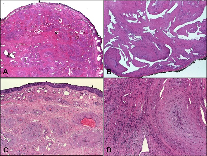
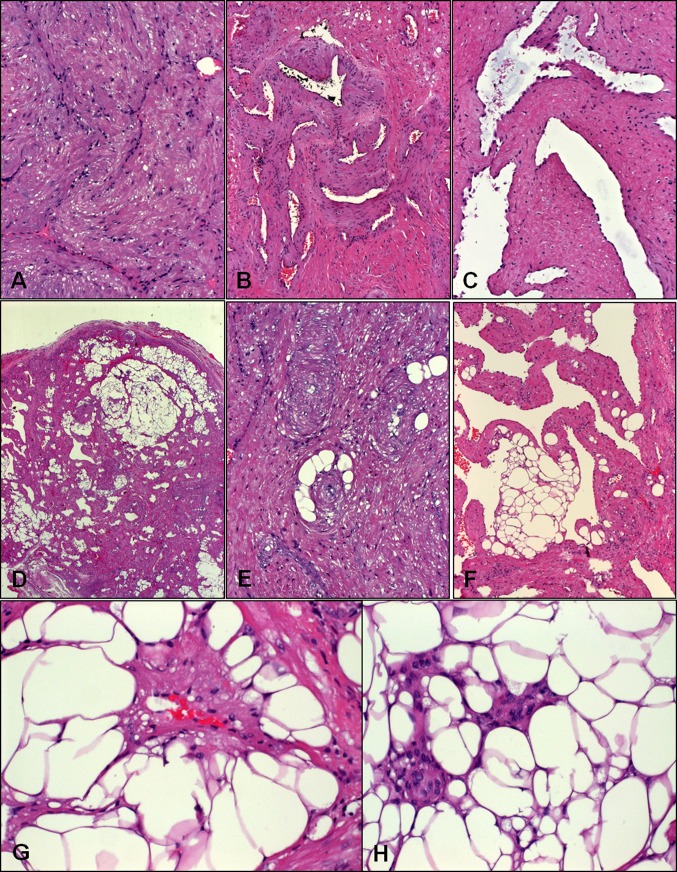
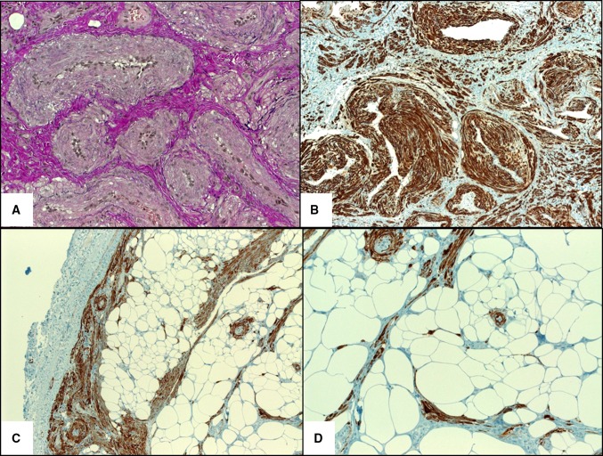
Angioleiomyoma (ALM; synonyms: angiomyoma, vascular leiomyoma) is an uncommon benign tumor of skin and subcutaneous tissue. Most arise in the extremities (90 %). Head and neck ALMs are uncommon (~10 % of all ALMs) and those arising beneath the sinonasal tract mucosa are very rare (<1 %) with 38 cases reported so far. We herein analyzed 16 cases identified from our routine and consultation files. Patients included seven females and nine males aged 25-82 years (mean 58; median 62). Symptoms were intermittent nasal obstruction, sinusitis, recurrent epistaxis, and a slow-growing mass. Fifteen lesions originated within different regions of the nasal cavity and one lesion was detected incidentally in an ethmoid sinus sample. Size range was 6-25 mm (mean 11). Histologically, all lesions were well circumscribed but non-encapsulated and most (12/16) were of the compact solid type superficially mimicking conventional leiomyoma but contained numerous compressed muscular veins. The remainder were of venous (2) and cavernous (2) type. Variable amounts of mature fat were observed in four cases (25 %). Atypia, necrosis, and mitotic activity were absent. Immunohistochemistry showed consistent expression of smooth muscle actin (12/12), h-caldesmon (9/9), muscle-specific actin (4/4), variable expression of desmin (11/14) and CD56 (4/6), and absence of HMB45 expression (0/11). The covering mucosa was ulcerated in 6 cases and showed squamous metaplasia in one case. There were no recurrences after local excision. Submucosal sinonasal ALMs are rare benign tumors similar to their reported cutaneous counterparts with frequent adipocytic differentiation. They should be distinguished from renal-type angiomyolipoma. Simple excision is curative.
Keywords: Angioleiomyoma; Angiomyolipoma; Angiomyoma; Nasal; PEComa; Sinonasal tract; Vascular leiomyoma.
Figures
References
-
- Fanburg-Smith JC, Thompson LDR. Benign soft tissue tumours. In: Barnes L, Eveson JW, Reichart P, Sidransky D, editors. World Health Organization classification of tumours. Pathology and genetics of head and neck tumours. Lyon: IARC Press; 2005. pp. 46–50.
Publication types
MeSH terms
Substances
LinkOut - more resources
Full Text Sources
Other Literature Sources
Medical
Research Materials
