Review
. 2016 May;90(5):1025-1048.
doi: 10.1007/s00204-015-1543-4. Epub 2015 Jun 6.

Experimental models of liver fibrosis

Affiliations
Review

Experimental models of liver fibrosis

Sara Crespo Yanguas et al. Arch Toxicol. 2016 May.

Abstract

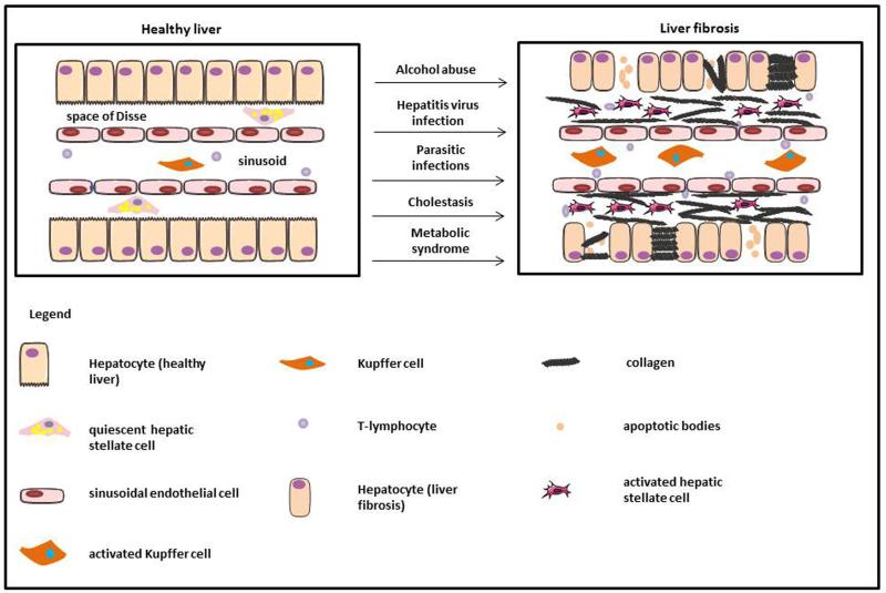
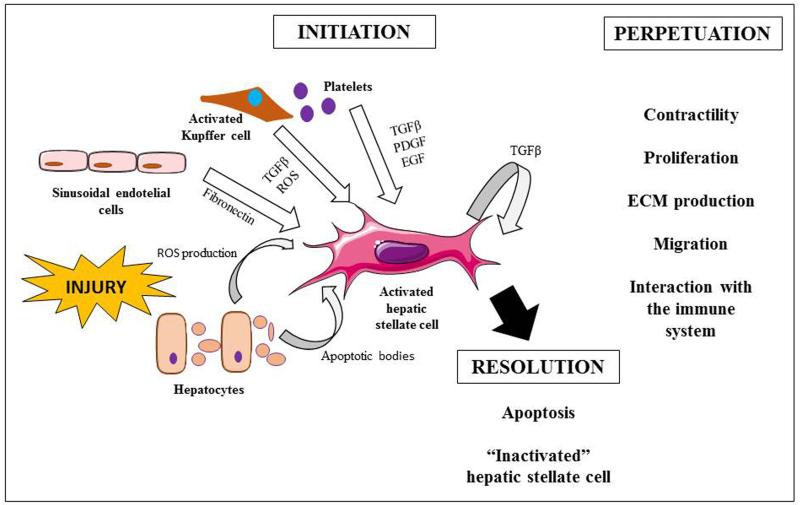
Hepatic fibrosis is a wound healing response to insults and as such affects the entire world population. In industrialized countries, the main causes of liver fibrosis include alcohol abuse, chronic hepatitis virus infection and non-alcoholic steatohepatitis. A central event in liver fibrosis is the activation of hepatic stellate cells, which is triggered by a plethora of signaling pathways. Liver fibrosis can progress into more severe stages, known as cirrhosis, when liver acini are substituted by nodules, and further to hepatocellular carcinoma. Considerable efforts are currently devoted to liver fibrosis research, not only with the goal of further elucidating the molecular mechanisms that drive this disease, but equally in view of establishing effective diagnostic and therapeutic strategies. The present paper provides a state-of-the-art overview of in vivo and in vitro models used in the field of experimental liver fibrosis research.

Keywords: Animal models; Hepatic stellate cells; In vitro models; Liver fibrosis.


Figures

Fig. 1
Fig. 1. Pathogenesis of liver fibrosis
In healthy liver, hepatocytes are studded with microvilli, HSCs store retinol and sinusoidal endothelial cells display fenestrae. During liver injury by a variety of causes, hepatocytes lose microvilli and may undergo apoptosis. The sinusoidal endothelial cells become devoid of fenestrae allowing inflammatory lymphocytes to infiltrate in the hepatic parenchyma. Furthermore Kupffer cells are activated, which in turn trigger HSC activation. As a result, large amounts of ECM proteins, including fibrillar collagens, are deposited in the Space of Disse.
Fig. 2
Fig. 2. Process of hepatic stellate cell activation
Upon insult, the stimuli that involve hepatic stellate cell (HSC) activation come from the injured hepatocytes, sinusoidal cells, Kupffer cells and platelets. Due to this interaction, HSCs are able to produce transforming growth factor β. The perpetuation of the injury leads to more active cells with the ability to contract, proliferate, produce extracellular matrix proteins, migrate and interact with the immune system. This triggers inflammatory and fibrogenic responses and decreases blood supply. Withdrawal of the injury may lead to the resolution of the disease by apoptosis of the activated HSCs and the reversion of the active into an inactivated HSC phenotype. (ECM, extracellular matrix; EGF, epithelial growth factor; PDGF, platelet derived growth factor; ROS, reactive oxygen species; TGFβ, transforming growth factor β)

References

    1. Abdel-Aziz G, Lebeau G, Rescan PY, et al. Reversibility of hepatic fibrosis in experimentally induced cholestasis in rat. Am J Pathol. 1990;137(6):1333–1342. - PMC - PubMed
    1. Abergel A, Sapin V, Dif N, et al. Growth arrest and decrease of alpha-SMA and type I collagen expression by palmitic acid in the rat hepatic stellate cell line PAV-1. Dig Dis Sci. 2006;51(5):986–995. - PubMed
    1. Abu-Absi SF, Hansen LK, Hu WS. Three-dimensional co-culture of hepatocytes and stellate cells. Cytotechnology. 2004;45(3):125–140. - PMC - PubMed
    1. Anstee QM, Goldin RD. Mouse models in non-alcoholic fatty liver disease and steatohepatitis research. Int J Exp Pathol. 2006;87(1):1–16. - PMC - PubMed
    1. Aparicio-Bautista DI, Pérez-Carreón JI, Gutiérrez-Nájera N, et al. Comparative proteomic analysis of thiol proteins in the liver after oxidative stress induced by diethylnitrosamine. Biochim Biophys Acta. 2013;1834(12):2528–2538. - PubMed

Publication types