

Review
doi: 10.1016/j.pbi.2015.05.007.
Epub 2015 Jun 3.
Refining the nuclear auxin response pathway through structural biology
Affiliations
- PMID: 26048079
- PMCID: PMC4618177
- DOI: 10.1016/j.pbi.2015.05.007
Item in Clipboard
Review
Refining the nuclear auxin response pathway through structural biology
Curr Opin Plant Biol.
2015 Oct.
Abstract
Auxin is a key regulator of plant growth and development. Classical molecular and genetic techniques employed over the past 20 years identified the major players in auxin-mediated gene expression and suggest a canonical auxin response pathway. In recent years, structural and biophysical studies clarified the molecular details of auxin perception, the recognition of DNA by auxin transcription factors, and the interaction of auxin transcription factors with repressor proteins. These studies refine the auxin signal transduction model and raise new questions that increase the complexity of auxin signaling.
Copyright © 2015 Elsevier Ltd. All rights reserved.
Figures
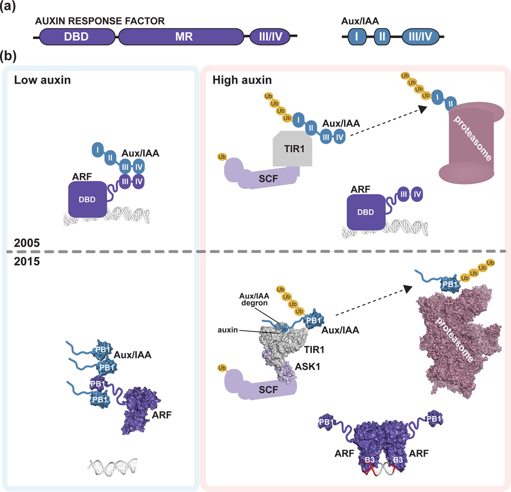
(a) Schematic of homology motifs in Auxin Response Factor (ARF) and Aux/IAA proteins. The ARF DNA binding domain (DBD), middle region (MR), and III/IV interaction motif are indicated. The Aux/IAA sequence motifs I (TOPLESS interaction), II (degron), and III/IV (interaction motif) are shown. (b) The top panel depicts the auxin response model from the year 2005. Under low local auxin concentrations, Aux/IAA proteins interact with ARF proteins, thereby repressing their action. In the presence of auxin , TIR1/AFB perceives auxin, allowing for SCFTIR1/AFB interaction with Aux/IAA proteins. Subsequently, Aux/IAA repressors are degraded via the 26S proteasome to allow for ARF-mediated gene expression changes. The bottom panel integrates structural biology contributions to our understanding of auxin signaling, including the ‘molecular glue’ interaction of auxin with TIR1 that allows for interaction of SCFTIR1/AFB with Aux/IAA repressors (PDB: 2P1Q; [22]); the ARF5 DBD structure (PDB: 4LDU; [34]) providing the ‘molecular calipers’ model of AuxRE recognition (PDB: 4LDX; [34]), and the possibility of PB1 domain multimerization in ARF (PDB: 4NJ6; [39]) and Aux/IAA (PDB: 2MUK; [42]) proteins to regulate auxin signaling. The cryo-EM proteasome structure is also shown for reference ([49]; PDB: 4CR2).
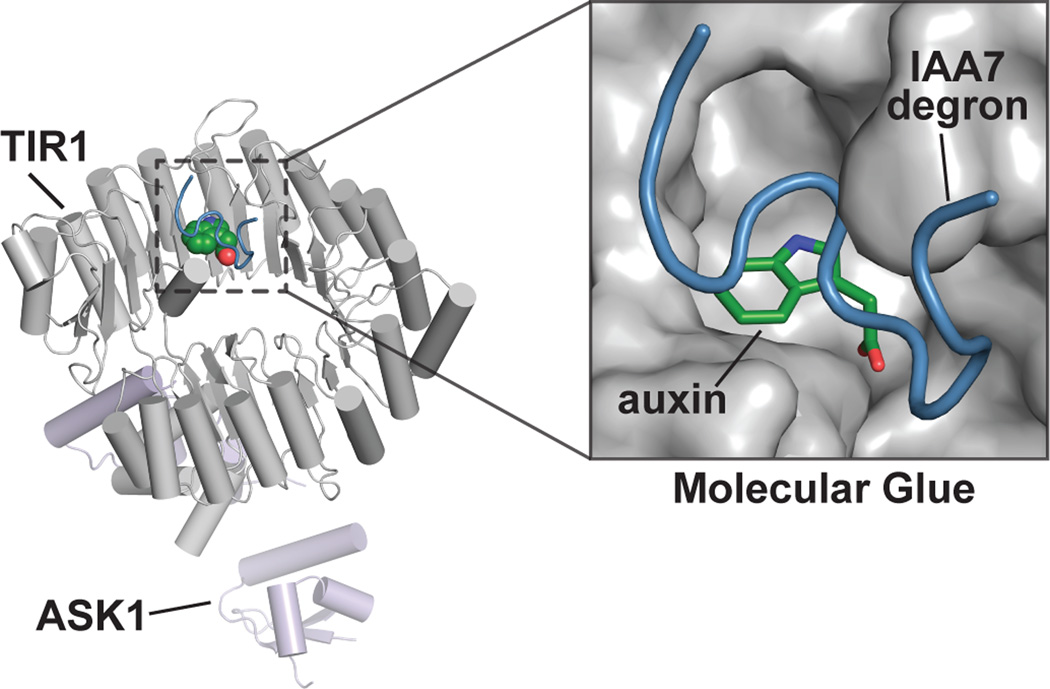
The x-ray crystal structure of the SCFTIR1/AFB•auxin•IAA7 peptide complex (PDB: 2P1Q; [22]) revels the presence of an auxin binding pocket that mediates Aux/IAA protein interaction. The overall structure of the complex formed by AtTIR1 (gray), AtASK1 (light purple), IAA (green), and a peptide corresponding to the AtIAA7 degron (blue) is shown. The inset depicts the TIR1 auxin-binding pocket, in which auxin acts as a molecular glue to allow interaction with Aux/IAA protein degron motifs.
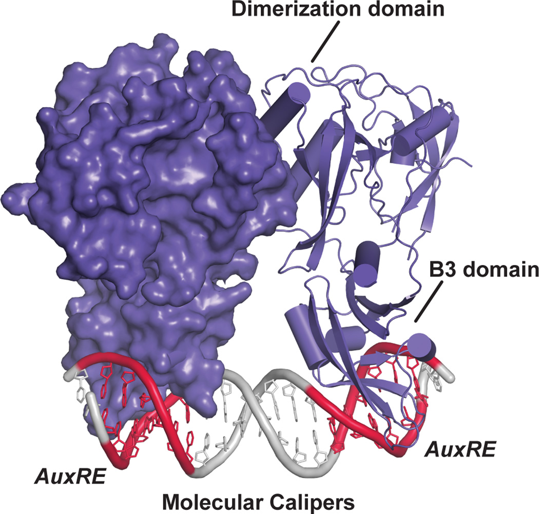
The structure of the AtARF1 dimerized DNA binding domain bound to the ER7 everted repeat containing two AuxREs (PDB: 4LDX; [34]) is shown. One monomer is shown as a surface rendering and the other as a ribbon diagram. The dimerization domain allows for spacing of the B3 DNA binding domain into the major groove of the DNA to interact with the AuxRE (red), leading to a suggested molecular calipers mechanism of DNA recognition by ARF proteins [34].
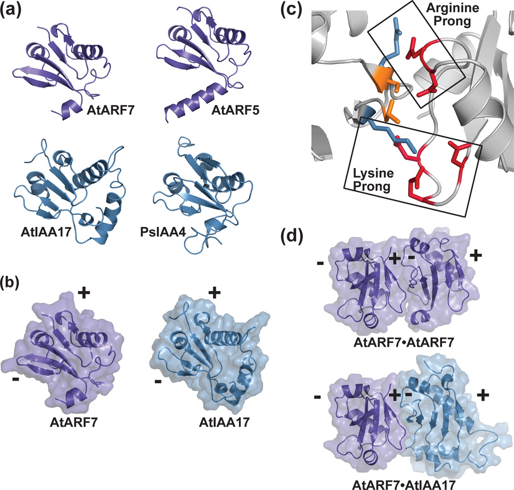
(a) The X-ray crystal structures of the PB1 domains of AtARF7 (PDB: 4NJ6; [39]) and AtARF5 (PDB: 4CHK; [40]) and the NMR solution structures of AtIAA17 (PDB: 2MUK; [42]) and PsIAA4 (PDB: 2MIM) reveal a common fold. (b) The PB1 domain scaffold presents conserved residues on opposing positive (+) and negative (-) interaction surfaces. A mixed surface/ribbon view of the PB1 domains of AtARF7 [39] and AtIAA17 [42] are shown. (c) PB1-mediated interaction of ARF and/or Aux/IAA proteins use a two-pronged binding motif. Biophysical studies of the ARF7 PB1 domain (PDB: 4NJ6; [39]) revealed how conserved lysine and arginine residues on the positive-face interact with two structurally distinct groups of acidic residues. (d) PB1 domains of ARF and Aux/IAA provide a versatile scaffold for protein interaction. Mixed surface/ribbon diagram showing the crystallographically-observed head-to-tail orientation of two AtARF7 PB1 domains (PDB: 4NJ6; [39]). Modeling of a hypothetical AtARF7•AtIAA17 complex, based on the AtARF7•AtARF7 structure, predicts the overall scaffold allowing for mixed-protein interaction.
References
-
- Peer WA. From perception to attenuation: auxin signalling and responses. Curr Opin Plant Biol. 2013;16:561–568. - PubMed
Publication types
MeSH terms
Substances
Grants and funding
LinkOut - more resources
Full Text Sources
Other Literature Sources
Miscellaneous
