. 2016 Jun;26(6):2705-14.
doi: 10.1093/cercor/bhv125. Epub 2015 Jun 5.

Brain Mechanisms for Processing Affective (and Nonaffective) Touch Are Atypical in Autism

Affiliations

Brain Mechanisms for Processing Affective (and Nonaffective) Touch Are Atypical in Autism

Martha D Kaiser et al. Cereb Cortex. 2016 Jun.

Abstract

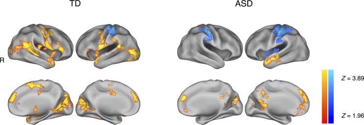
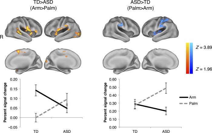
C-tactile (CT) afferents encode caress-like touch that supports social-emotional development, and stimulation of the CT system engages the insula and cortical circuitry involved in social-emotional processing. Very few neuroimaging studies have investigated the neural mechanisms of touch processing in people with autism spectrum disorder (ASD), who often exhibit atypical responses to touch. Using functional magnetic resonance imaging, we evaluated the hypothesis that children and adolescents with ASD would exhibit atypical brain responses to CT-targeted touch. Children and adolescents with ASD, relative to typically developing (TD) participants, exhibited reduced activity in response to CT-targeted (arm) versus non-CT-targeted (palm) touch in a network of brain regions known to be involved in social-emotional information processing including bilateral insula and insular operculum, the right posterior superior temporal sulcus, bilateral temporoparietal junction extending into the inferior parietal lobule, right fusiform gyrus, right amygdala, and bilateral ventrolateral prefrontal cortex including the inferior frontal and precentral gyri, suggesting atypical social brain hypoactivation. Individuals with ASD (vs. TD) showed an enhanced response to non-CT-targeted versus CT-targeted touch in the primary somatosensory cortex, suggesting atypical sensory cortical hyper-reactivity.

Keywords: affective touch; autism spectrum disorder; functional magnetic resonance imaging; insula; sensory hyper-reactivity; tactile perception.


Figures

Figure 1.
Figure 1.
Whole-brain results from a contrast of (Arm > Palm) (orange-to-yellow color map) and (Palm > Arm) (blue-to-light blue). The TD group is represented on the left and the ASD group on the right. Left-hand panel: TD children and adolescents exhibited Arm > Palm (orange-to-yellow color map) activity in bilateral insular cortex extending into the ventrolateral prefrontal cortices (vlPFC) and the temporal poles as well as the right posterior superior temporal sulcus (pSTS), right amygdala, and right fusiform gyrus (FG). Palm > Arm activity (blue-to-light blue color map) was localized to the expected location of the primary somatosensory cortex (S1). While the children and adolescents with ASD shared Palm > Arm activity in the S1 (right-hand panel), they lacked Arm > Palm activity in the right pSTS, bilateral temporoparietal junction (TPJ) extending into the inferior parietal lobule (IPL), right FG, right amygdala, bilateral insula, and vlPFC, identified in TD participants. We controlled for age as a covariate of no interest. These results were estimated using FSL's mixed-effects algorithm (FLAME1 + 2) and corrected for multiple comparisons at a voxel-level threshold Z> 1.96, cluster-level threshold P < 0.05.
Figure 2.
Figure 2.
Whole-brain results from contrasts of TD > ASD on (Arm > Palm) and ASD > TD on (Palm > Arm). Evaluation of the Group × Condition interaction identified regions where the ASD and TD groups displayed distinct differential responses to the (Arm > Palm) and (Palm > Arm) contrasts. Top, left: The TD group uniquely exhibited an enhanced response to CT-targeted Arm > Palm touch (orange-to-yellow color map) including the bilateral anterior insula and insular operculum extending into vlPFC, right pSTS, bilateral TPJ (extending into the IPL), and right FG. There were no significantly greater activations in the ASD (Arm > Palm) > TD (Arm > Palm) contrast. Top, right: The ASD group uniquely exhibited an enhanced response to non-CT-targeted Palm > Arm touch (blue-to-light blue color map) including primarily the S1. Age was controlled for as a covariate of no interest. The results were estimated using FSL's mixed-effects algorithm (FLAME1 + 2) and corrected for multiple comparisons at a voxel-level threshold Z > 1.96, cluster-level threshold P < 0.05. Bottom: Percent signal change of the Arm and Palm conditions versus the baseline of rest periods by group (TD and ASD) and on regions corresponding to the left and right top panels, respectively. Errors bars represent standard errors.
Figure 3.
Figure 3.
Percent signal change analyses of the (Arm or Palm) conditions versus the baseline of rest periods by group (TD or ASD). We used anatomically defined right insular cortex (top, outlined in blue) and right pSTS (bottom, outlined in yellow) as region of interests (ROIs). In both ROIs, it is primarily Arm (vs. rests), but not Palm (vs. rests), that drives the TD > ASD difference. Errors bars represent standard errors.
Figure 4.
Figure 4.
Functional connectivity analyses of TD > ASD on (Arm > Palm). We used anatomically defined right insular cortex (top, outlined in blue) and right pSTS (bottom, outlined in yellow) as seeds. Top: in TD relative to ASD, greater functional connectivity was identified between (A) the right insula and ventromedial prefrontal cortex (vmPFC), and (B) the right insula and the paracentral lobule. Bottom: in TD relative to ASD, greater functional connectivity was identified between (A) the right pSTS and the right amygdala extending into the hippocampus and (B) the right pSTS and the anterior temporal lobe, extending into the FG. Age was controlled for as a covariate of no interest. The results were estimated using FSL's mixed-effects algorithm (FLAME1 + 2) and corrected for multiple comparisons at a voxel-level threshold Z > 1.96, cluster-level threshold P < 0.05.
Figure 5.
Figure 5.
Results of a whole-brain correlation analysis between ADOS Calibrated Severity Scores (CSS) and Arm versus Palm activities. (A) Greater levels of autism symptom severity were associated with decreased reactivity to (Arm > Palm) in the left precentral gyrus and the left postcentral gyrus (orange-to-yellow color map). (B) Greater levels of autism symptom severity were associated with decreased reactivity to (Arm + Palm) in brain regions including primarily the medial prefrontal cortex (green-to-light green color map). Age was controlled for as a covariate of no interest. The results were estimated using FSL's mixed-effects algorithm (FLAME1 + 2) and corrected for multiple comparisons at a voxel-level threshold Z > 1.96, cluster-level threshold P < 0.05. **P < 0.01.

References

    1. Abrams DA, Lynch CJ, Cheng CM, Phillips J, Supekar K, Ryali S, Uddin LQ, Menon V. 2013. Underconnectivity between voice-selective cortex and reward circuitry in children with autism. Proc Natl Acad Sci USA. 110:12061–12065. - PMC - PubMed
    1. American Psychiatric Association. 2013. Diagnostic and Statistical Manual of Mental Disorders. 5th ed Arlington: (VA: ): American Psychiatric Publishing.
    1. American Academy of Pediatrics. 2012. Sensory integration therapies for children with developmental and behavioral disorders. Pediatrics. 129:1186–1189. - PubMed
    1. Baranek GT. 1999. Autism during infancy: a retrospective video analysis of sensory-motor and social behaviors at 9–12 months of age. J Autism Dev Disord. 29:213–224. - PubMed
    1. Barnett L. 2005. Keep in touch: the importance of touch in infant development. Infant Obs. 8:115–123.

Publication types