

Functional Organization of Social Perception and Cognition in the Superior Temporal Sulcus
- PMID: 26048954
- PMCID: PMC4816802
- DOI: 10.1093/cercor/bhv111
Functional Organization of Social Perception and Cognition in the Superior Temporal Sulcus
Abstract
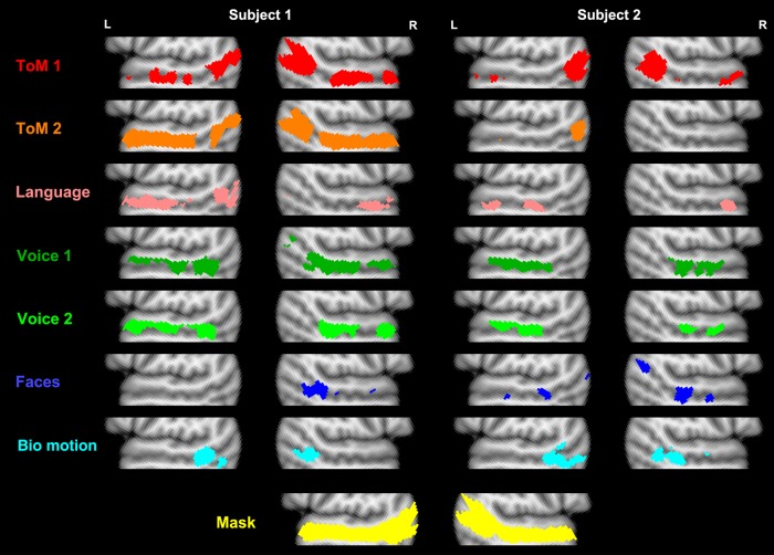
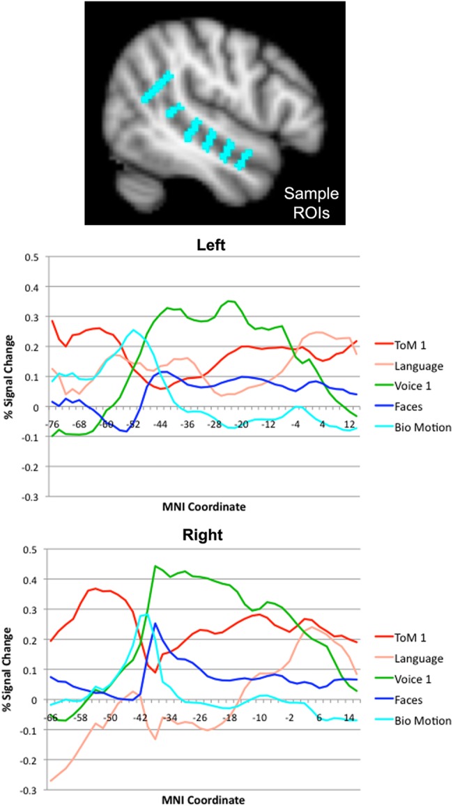
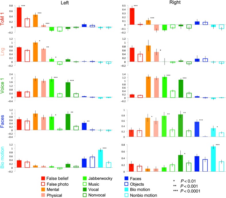
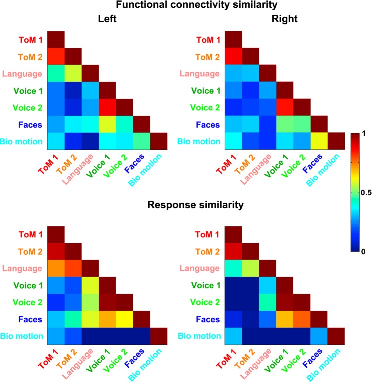
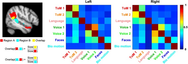
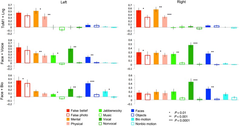
The superior temporal sulcus (STS) is considered a hub for social perception and cognition, including the perception of faces and human motion, as well as understanding others' actions, mental states, and language. However, the functional organization of the STS remains debated: Is this broad region composed of multiple functionally distinct modules, each specialized for a different process, or are STS subregions multifunctional, contributing to multiple processes? Is the STS spatially organized, and if so, what are the dominant features of this organization? We address these questions by measuring STS responses to a range of social and linguistic stimuli in the same set of human participants, using fMRI. We find a number of STS subregions that respond selectively to certain types of social input, organized along a posterior-to-anterior axis. We also identify regions of overlapping response to multiple contrasts, including regions responsive to both language and theory of mind, faces and voices, and faces and biological motion. Thus, the human STS contains both relatively domain-specific areas, and regions that respond to multiple types of social information.
Keywords: social cognition; social perception; superior temporal sulcus.
© The Author 2015. Published by Oxford University Press.
Figures
Comment in
-
The social mysteries of the superior temporal sulcus.Trends Cogn Sci. 2015 Sep;19(9):489-90. doi: 10.1016/j.tics.2015.07.002. Epub 2015 Jul 21. Trends Cogn Sci. 2015. PMID: 26208834 Free PMC article.
References
-
- Allison T, Puce A, McCarthy G. 2000. Social perception from visual cues: role of the STS region. Trends Cogn Sci. 4:267–278. - PubMed
-
- Ambady N, Rosenthal R. 1992. Thin slices of expressive behavior as predictors of interpersonal consequences: a meta-analysis. Psychol Bull. 111:256.
-
- Apperly IA, Samson D, Carroll N, Hussain S, Humphreys G. 2006. Intact first-and second-order false belief reasoning in a patient with severely impaired grammar. Soc Neurosci. 1:334–348. - PubMed
-
- Apperly IA, Samson D, Chiavarino C, Humphreys GW. 2004. Frontal and temporo-parietal lobe contributions to theory of mind: neuropsychological evidence from a false-belief task with reduced language and executive demands. J Cogn Neurosci. 16:1773–1784. - PubMed
-
- Barraclough NE, Xiao D, Baker CI, Oram MW, Perrett DI. 2005. Integration of visual and auditory information by superior temporal sulcus neurons responsive to the sight of actions. J Cogn Neurosci. 17:377–391. - PubMed
Publication types
MeSH terms
Substances
LinkOut - more resources
Full Text Sources
Other Literature Sources
