Review
. 2015 Jul;25(3):155-63.
doi: 10.1016/j.semradonc.2015.02.006. Epub 2015 Feb 23.

Molecular Markers in Low-Grade Glioma-Toward Tumor Reclassification

Affiliations
Review

Molecular Markers in Low-Grade Glioma-Toward Tumor Reclassification

Adriana Olar et al. Semin Radiat Oncol. 2015 Jul.

Abstract

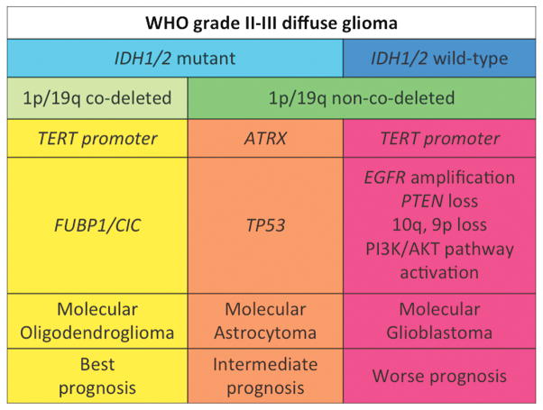
Low-grade diffuse gliomas are a heterogeneous group of primary glial brain tumors with highly variable survival. Currently, patients with low-grade diffuse gliomas are stratified into risk subgroups by subjective histopathologic criteria with significant interobserver variability. Several key molecular signatures have emerged as diagnostic, prognostic, and predictor biomarkers for tumor classification and patient risk stratification. In this review, we discuss the effect of the most critical molecular alterations described in diffuse (IDH1/2, 1p/19q codeletion, ATRX, TERT, CIC, and FUBP1) and circumscribed (BRAF-KIAA1549, BRAF(V600E), and C11orf95-RELA fusion) gliomas. These molecular features reflect tumor heterogeneity and have specific associations with patient outcome that determine appropriate patient management. This has led to an important, fundamental shift toward developing a molecular classification of World Health Organization grade II-III diffuse glioma.


Conflict of interest statement

Conflict of interest: none.

Figures

Figure 1
Figure 1
Diffuse gliomas are aggressive neoplasms that tend to infiltrate along dendrites and axons and travel long distances from the primary site of origin. Malignant glioma cells are shown infiltrating the cortex and forming aggregates along a blood vessel (yellow arrows) extending all the way to the leptomeningeal surface (black arrows) (A – H&E, Obj: 100X). A higher power of the cortex highlights neoplastic glioma cells (green arrow) surrounding the neurons (yellow arrow) (B – H&E, Obj: 200X). A special immunohistochemical stain for the mutated IDH-R132H protein highlights (in brown) malignant glioma cells infiltrating the normal (pale) subcortical and cortical tissue – note how the subcortex has an incresed density of glioma cells (left upper corner) compared to the superficial cortex (right lower corner) (C – IDH1-R132H, Obj: 40X, scale not available).
Figure 2
Figure 2
Diffuse glioma with 1p/19q co-deletion. The locus-specific identifier (LSI) probe, 1p36 (SpectrumOrange) and corresponding LSI 1q25 (SpectrumGreen) show two normal green signals and single abnormal orange signal in a subpopulation of interphase cells suggestive of 1p loss (left). LSI probe, 19q13 (SpectrumOrange) and corresponding LSI 19p13 (SpectrumGreen) show two normal green signals and single abnormal orange signal in a subpopulation of oligodendroglioma interphase cells suggestive of 19q loss (right) (Photographs courtesy of Prof. Dr. Adekunle Adesina, Texas Children’s Hospital, Houston, TX, USA).
Figure 3
Figure 3
Proposed molecular classification of WHO grade II-III diffuse glioma.

References

    1. Ostrom QT, Gittleman H, Liao P, et al. CBTRUS Statistical Report: Primary Brain and Central Nervous System Tumors Diagnosed in the United States in 2007–2011. Neuro-oncology. 2014;16(Suppl 4):iv1–iv63. - PMC - PubMed
    1. Louis DN, Ohgaki H, Wiestler OD, et al. WHO Classification of tumors of the central nervous system. Lyon (France): IARC; 2007. - PMC - PubMed
    1. van den Bent MJ. Interobserver variation of the histopathological diagnosis in clinical trials on glioma: a clinician’s perspective. Acta Neuropathol. 2010;120(3):297–304. - PMC - PubMed
    1. Aldape K, Burger PC, Perry A. Clinicopathologic aspects of 1p/19q loss and the diagnosis of oligodendroglioma. Arch Pathol Lab Med. 2007;131(2):242–51. - PubMed
    1. Coons SW, Johnson PC, Scheithauer BW, et al. Improving diagnostic accuracy and interobserver concordance in the classification and grading of primary gliomas. Cancer. 1997;79(7):1381–93. - PubMed

Publication types

Substances