. 2015 Jun 17;86(6):1385-92.
doi: 10.1016/j.neuron.2015.05.022. Epub 2015 Jun 4.

Motor Learning Consolidates Arc-Expressing Neuronal Ensembles in Secondary Motor Cortex

Affiliations

Motor Learning Consolidates Arc-Expressing Neuronal Ensembles in Secondary Motor Cortex

Vania Yu Cao et al. Neuron. .

Abstract

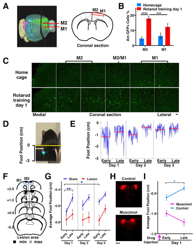
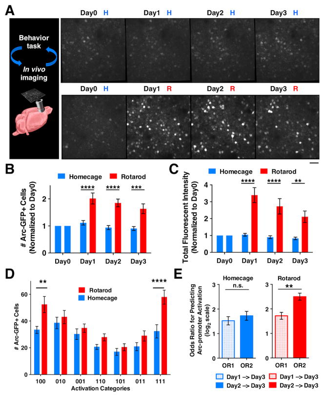
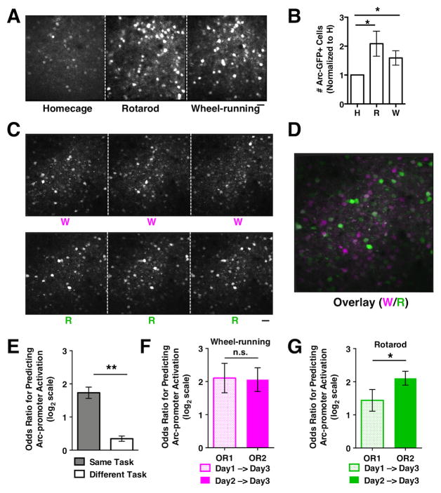
Motor behaviors recruit task-specific neuronal ensembles in motor cortices, which are consolidated over subsequent learning. However, little is known about the molecules that can identify the participating neurons and predict the outcomes of the consolidation process. Using a mouse rotarod-learning task, we showed that lesion or inactivation of the secondary motor (M2) cortex disrupts learning of skilled movements. We tracked the endogenous promoter activity of the neuronal activity-regulated gene Arc in individual M2 neurons during rotarod learning by in vivo two-photon imaging of a knockin reporter. We found that task training initially recruits Arc-promoter-activated neurons and then consolidates them into a specific ensemble exhibiting persistent reactivation of Arc-promoter. The intensity of a neuron's initial Arc-promoter activation predicts its reactivation probability and neurons with weak initial Arc-promoter activation are dismissed from the ensemble during subsequent training. Our findings demonstrate a task-specific Arc-dependent cellular consolidation process in M2 cortex during motor learning.


Figures

Figure 1
Figure 1. Rotarod training recruits neurons in M2 cortex and M2 function is required for learning skilled stepping movements
(A) A 3D mouse brain model (Allen Brain Atlas) and coronal section schematic. The red rectangle indicates superficial layers of the motor cortical regions imaged in B and C. (B) Preferential activation of Arc-GFP by rotarod training in M2 compared to M1 (two-way RM-ANOVA, region-by-behavior, F(1,10)=23.36, P=0.0007; Rotarod M2 vs. M1, P=0.0008; M2 Rotarod vs. Homecage, P<0.0001; N=6 mice per condition). The percentage of GFP+ cells was estimated from the total number of GFP+ (green) and GFP− (dark) cells. (C) Confocal image montage of coronal sections from heterozygous Arc-GFP mice. Scale bars, 30 μm. (D) A video frame showing mouse performing the accelerating rotarod task. The distance from the left rear paw of the mouse (blue star) to the apex of the rod (yellow line) was measured. (E) Foot position traces (blue) of a normal mouse during Early and Late trials from 3 training days. The red lines indicate the average foot position in each trial. (F) Illustration of minimum and maximum extents of M2 surgical lesions: N=23 mice. (G) Average foot positions during Early and Late trials from sham and M2 lesion groups. Two-way RM ANOVA, lesion effect, F(1,44)=8.86, P=0.0047, N=23 mice per group. Short-term learning, Early vs. Late on Day 1: Sham P=0.0057, Lesion P=0.46. Long-term learning, Day 1 vs. Day 3 Early: Sham P=0.014, Lesion P=0.80. (H) Confocal images of coronal sections showing injection sites for muscimol inactivation of M2 cortex. Dextran-TRITC (red) was mixed in ACSF and injected into the M2 cortex in the presence or absence of muscimol (0.5μl, 1μg/μl) 45 min before rotarod training. Scale bar, 500 μm. (I) Average foot positions during Early and Late trials on training Day 1 with muscimol inactivation. Two-way RM ANOVA, muscimol effect, F(1,15)=61.07, P<0.0001, N=11 mice for control and 10 mice for muscimol group. Early vs. Late: Control P=0.013, Muscimol P=0.11. Error bars indicate SEM.
Figure 2
Figure 2. M2 neuronal ensembles identified by Arc-promoter activation are consolidated in subsequent days of rotarod training
(A) In vivo two-photon images showing Arc-GFP expression in superficial layer (~180 μm from pia) M2 neurons of a heterozygous Arc-GFP mouse under the repeated homecage (H) condition, and another mouse under the repeated rotarod (R) training condition. Scale bar 30μm. (B) The number of Arc-GFP+ neurons (normalized to the level on Day 0) in homecage (N=11) and rotarod trained (N=10) mice. Two-way RM ANOVA, rotarod effect, F(1,19)=28.92, P<0.0001; H vs R: Day 1 P<0.0001; Day 2 P<0.0001, Day 3 P=0.0002. (C) Total fluorescence intensity of matched cell regions (normalized to the level on Day 0) in homecage and rotarod trained mice. Two-way RM ANOVA, rotarod effect, F(1,19)=25.66, P<0.0001; H vs R: Day 1 P<0.0001, Day 2 P<0.0001, Day 3 P=0.0025. (D) The number of Arc-GFP+ neurons in each of the 3-day activation categories under the Homecage (HHH) and Rotarod (RRR) conditions. Two-way RM ANOVA, condition-by-category, F(6,174)=2.985, P=0.0084; H vs. R: “100”, P=0.004; “111”, P<0.0001). (E) Odds ratios (OR) for predicting Arc-promoter activation. Under the homecage condition, day-1 (OR1) and day-2 (OR2) Arc-promoter activation have similar effectiveness in predicting the reactivation on day 3 (paired t-test, P=0.280, N=16 mice). Under the rotarod condition, day-2 Arc-promoter activation is more effective than day-1 activation in predicting the reactivation on day 3 (paired t-test, P=0.0063, N=15 mice). Error bars indicate SEM.
Figure 3
Figure 3. Task-specific recruitment and consolidation of neuronal ensembles defined by Arc-promoter activation
(A) In vivo two-photon images showing Arc-GFP expression in superficial layer M2 neurons of a mouse under consecutive days of homecage (H), rotarod (R), and wheel-running (W) conditions. (B) The numbers of Arc-GFP+ cells detected under each condition (normalized to H, paired t-test, R vs. H, P=0.040; W vs. H, P=0.046, N=9 mice). (C) In vivo two-photon images showing Arc-GFP expression in M2 neurons of a mouse under the repeated wheel-running (W) and rotarod-training (R) conditions. (D) Overlay of M2 images under the wheel-running (magenta) and rotarod-training (green) conditions. Cells that are preferentially activated in either condition appear as magenta or green, whereas cells that are equally activated by both conditions appear as white. (E) The odds ratio for predicting Arc-promoter activation under the same motor task (W-W, R-R) is significantly higher than that between the different motor tasks (W-R). Paired t-test, P=0.0012, N=5 mice. (F) Under the wheel-running condition, day-1 (OR1) and day-2 (OR2) Arc-promoter activation has similar effectiveness in predicting the reactivation on day 3 (paired t-test, P=0.924, N=5 mice). (G) Under the rotarod condition, day-2 Arc-promoter activation is more effective than day-1 activation in predicting the reactivation on day 3 (paired t-test P=0.042, N=5 mice). Scale bars, 30μm. Error bars indicate SEM.
Figure 4
Figure 4. The initial intensity of Arc-promoter activation in a neuron predicts its probability of dismissal or retention in the rotarod ensemble
(A) Close-up images of individual M2 neurons showing their fluorescent intensity on rotarod training Day 1 and the classification of their reactivation categories on Day 2 and 3. Red arrowheads indicate additional “Retained” neurons that are near “Dismissed” or “Unstable” neurons. Scale bar, 10 μm. (B) The reactivation category probabilities are dependent on the fluorescent intensity of Day 1 Arc-GFP expression in cells (Chi-square test, P<0.0001, 2376 neurons from 15 mice). (C) Neurons with relatively weak initial Arc-promoter activation are more likely to be dismissed from the rotarod ensemble, whereas neurons with relatively strong initial Arc-promoter activation are more likely to be retained. (D) Diagram showing that the initial intensity of Arc-promoter activation in a neuron predicts its probability of dismissal or retention during the cellular consolidation process.

References

    1. Bramham CR, Worley PF, Moore MJ, Guzowski JF. The immediate early gene arc/arg3.1: regulation, mechanisms, and function. J Neurosci. 2008;28:11760–11767. - PMC - PubMed
    1. Brecht M. Movement, confusion, and orienting in frontal cortices. Neuron. 2011;72:193–196. - PubMed
    1. Buitrago MM, Schulz JB, Dichgans J, Luft AR. Short and long-term motor skill learning in an accelerated rotarod training paradigm. Neurobiol Learn Mem. 2004;81:211–216. - PubMed
    1. Cao VY, Ye Y, Mastwal SS, Lovinger DM, Costa RM, Wang KH. In vivo two-photon imaging of experience-dependent molecular changes in cortical neurons. Journal of visualized experiments: JoVE 2013 - PMC - PubMed
    1. Costa RM, Cohen D, Nicolelis MA. Differential corticostriatal plasticity during fast and slow motor skill learning in mice. Curr Biol. 2004;14:1124–1134. - PubMed

Publication types

Substances

LinkOut - more resources