. 2015 Jun 22;33(6):729-36.
doi: 10.1016/j.devcel.2015.05.004. Epub 2015 Jun 4.

Mechanical Allostery: Evidence for a Force Requirement in the Proteolytic Activation of Notch

Affiliations

Mechanical Allostery: Evidence for a Force Requirement in the Proteolytic Activation of Notch

Wendy R Gordon et al. Dev Cell. .

Abstract

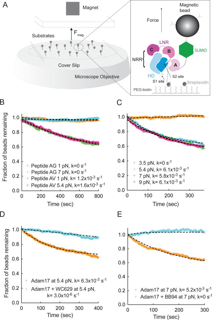
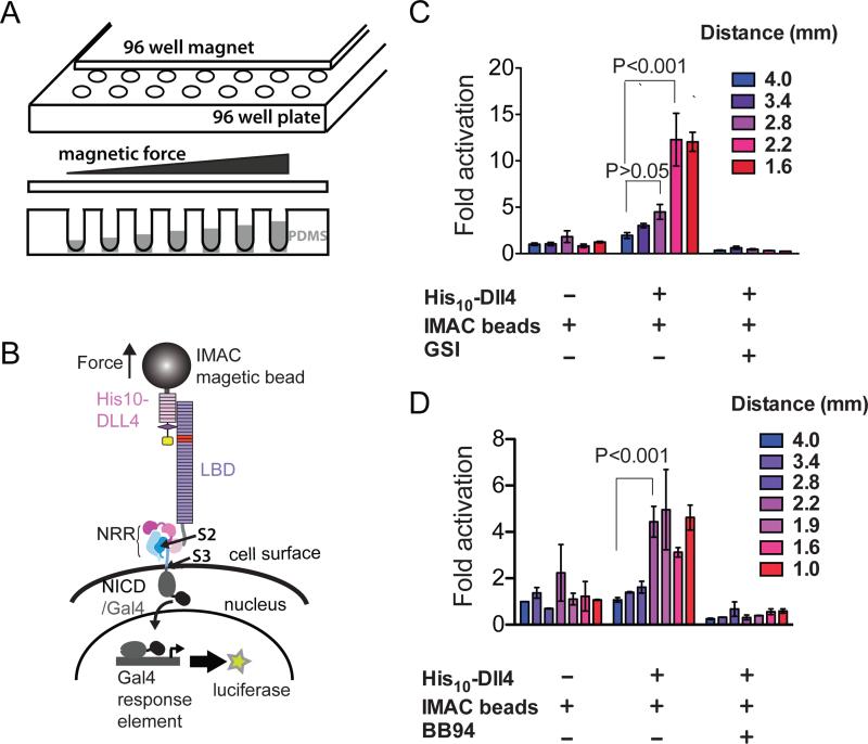
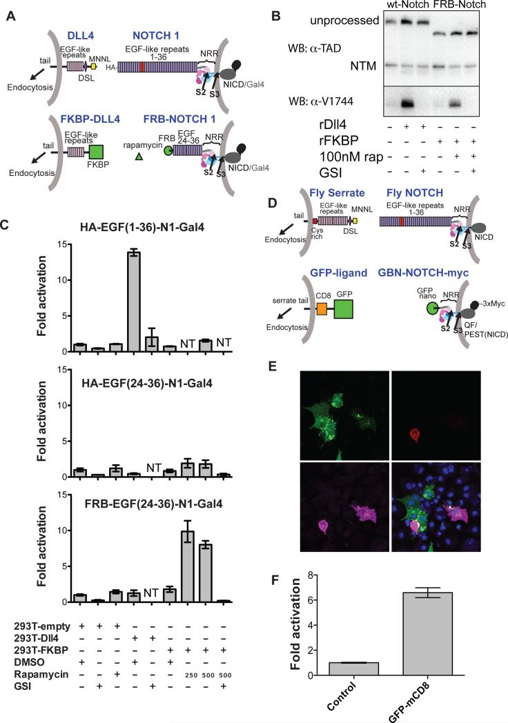
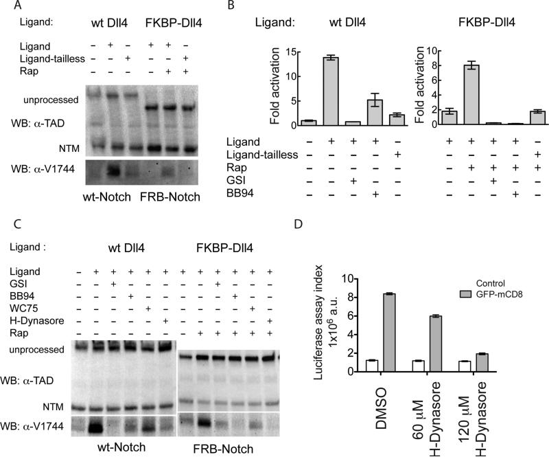
Ligands stimulate Notch receptors by inducing regulated intramembrane proteolysis (RIP) to produce a transcriptional effector. Notch activation requires unmasking of a metalloprotease cleavage site remote from the site of ligand binding, raising the question of how proteolytic sensitivity is achieved. Here, we show that application of physiologically relevant forces to the Notch1 regulatory switch results in sensitivity to metalloprotease cleavage, and bound ligands induce Notch signal transduction in cells only in the presence of applied mechanical force. Synthetic receptor-ligand systems that remove the native ligand-receptor interaction also activate Notch by inducing proteolysis of the regulatory switch. Together, these studies show that mechanical force exerted by signal-sending cells is required for ligand-induced Notch activation and establish that force-induced proteolysis can act as a mechanism of cellular mechanotransduction.


Figures

Figure 1
Figure 1. Single molecule assay of Adam17-mediated proteolysis of the Notch1 NRR under force
(A) Assay schematic and experimental design. Magnetic beads are tethered to proteins immobilized in a flowcell mounted on an inverted microscope. Force is applied to the beads by varying the distance between a magnet and the surface of the flowcell. Substrate proteolysis is monitored by determining the fraction of beads released over time. The expanded view in the right-hand panel illustrates the Notch1 NRR, captured on the flow cell with streptavidin and tethered to the magnetic bead using anti-SUMO antibodies. (B) Adam17-catalyzed proteolysis of biotinylated and SUMO tagged recombinant peptides, containing either the natural S2-cleavage site sequence (AV, green and pink), or a mutated sequence with a V1721G substitution (AG, orange and cyan) at the forces indicated. (C) Adam17-catalyzed proteolysis of the Notch1 NRR, monitored as a function of time at different levels of applied force. Traces shown represent averages of 2 or 3 replicates. (D, E) Effect of inhibitors on proteolysis of the Notch1 NRR in the single molecule cleavage assay. Traces shown represent a single experiment. (D) Effect of WC629, an anti-Notch1 inhibitory antibody that binds to the NRR, on the time course of Adam17-catalyzed NRR proteolysis. (E) Effect of BB94, an ADAM inhibitor, on the time course of Adam17-catalyzed NRR proteolysis. Additional control experiments are provided in Figure S1.
Figure 2
Figure 2. Response of cell-surface Notch receptors to applied force using a multiplexed magnetic tweezers assay
(A) Experimental design. A plate containing 96 cylindrical magnets is positioned over a 96 well plate of cells in order to apply force to magnetic beads tethered to Notch molecules on the cell-surface. The distance between the cells and the magnet is varied by using the polymer PDMS to create terraces of different heights. (B) Assay schematic. Cells expressing Notch1 receptors in which the ankyrin-repeat domain has been replaced by the Gal4 DNA-binding domain (Malecki et al., 2006) are stimulated by magnetic beads loaded with the ligand DLL4, followed by measurement of luciferase reporter gene activity. (C, D) Luciferase reporter gene activity in response to various treatments as a function of the distance from the magnet. U2OS cells expressing Notch1-Gal4 receptors were incubated with magnetic beads alone or beads loaded with the ligand DLL4 in the absence or presence of a gamma secretase inhibitor (GSI) (C) or metalloprotease inhibitor BB94 (D). Luciferase reporter gene activity is reported relative to the response of cells to beads alone at a distance of 4 mm from the magnet. Error bars represent the standard error of the mean of triplicate measurements, and statistical significance was determined with a two-way ANOVA followed by a post-hoc Bonferroni's test. 96-well magnet calibration is provided in Figure S2.
Figure 3
Figure 3. Development and evaluation of two synthetic Notch signaling systems
(A) Schematic comparing the natural human Notch-ligand signaling system (top; EGF repeats 11-12 in red) to a synthetic signaling system placing NRR proteolysis under rapamycin-inducible control (bottom). Here, FKBP replaces the N-terminal portion of DLL4, and FRB replaces EGF-like repeats 1-23 of Notch1. The Notch1 ankyrin domain is also replaced with Gal4, as above (Malecki et al., 2006). (B) Western blots monitoring receptor proteolysis. U2OS cells stably expressing wild-type or FRB-Notch1 were grown in the presence of the DLL4 ectodomain or FKBP immobilized on plastic tissue culture dishes in the absence or presence of rapamycin (250 nM) and/or a GSI (Compound E, 400 nM). Blots were probed with an antibody directed against an epitope of intracellular Notch1 (α-TAD), or the α-V1744 antibody to S3-cleaved Notch1 (Cell Signaling). (C) Cell-based reporter gene assay. U2OS cells stably transfected with the indicated Notch variants were co-cultured with 293T cells transiently transfected with the indicated ligands. Luciferase activity for each U2OS line is reported relative to co-culture with 293T cells transfected with empty vector. Error bars reflect the standard error of readings performed in triplicate. Additional control experiments are provided in Figure S3. (D) Schematic illustrating design of a GFP - GFP-binding nanobody (GBN) synthetic ligand-receptor pair. Full-length fly Serrate and Notch are shown for reference. The artificial ligand consists of GFP, CD8 and the Serrate-derived tail. The ectodomain of the Notch-derived molecule consists of the GFP binding nanobody (GBN) and the NRR, and the intracellular domain contains the QF transcription factor, the Notch PEST domain, and a triple Myc tag. (E) Co-culture assay. S2R+ cells expressing GFP-mcd8-Ser as ligand (green, upper left panel) were co-cultured with cells expressing GBN-FlyNotch(NRR)-QF-3XMyc (GBN-N-QFMyc). Receptor is stained with anti-myc antibody (magenta, lower left panel). The tdTomato reporter signal is red (upper right panel). DNA was stained with DAPI (blue, lower right panel). (F) QAS luciferase readout of cell-mixing experiment. Luciferase reporter gene activity for the GBN-Notch cell line is reported relative to co-culture with control cells. Error bars represent the standard error of measurements performed in quadruplicate. Additional supporting data are provided in Figure S3.
Figure 4
Figure 4
Western blot analysis of natural and synthetic Notch receptor signaling in co-culture assays. (A) Effect of ligand-tail deletion on signaling. Wild-type and synthetic Notch receptors were co-cultured with full-length or tail-deleted cognate ligands (Ligand-tailless) and in the absence or presence of rapamycin (250 nM), as indicated. Blots were probed with an antibody directed against an epitope of intracellular Notch1 (α-TAD), or the α-V1744 antibody to S3-cleaved Notch1 (Cell Signaling). (B) Cell-based reporter gene assay probing Notch activation in co-culture experiments. 293T cells were signal-sending cells, and U2OS cells were signal-receiving cells. 293T cells were transfected with plasmids encoding wild-type Dll4, tailless Dll4, FKBP-Dll4, FKBP-Dll4-tailless or empty vector in the presence or absence of rapamycin (250 nM), Compound E (GSI, 400nM), or BB94 (20 μM). U2OS cells were transfected in 96-well format with plasmids encoding HA-Notch1-Gal4 (left), or FRB-Notch1-Gal4 (right) along with a luciferase reporter plasmid containing the Gal4 response element and an internal control plasmid expressing Renilla luciferase. 24 h after transfection, the 293T cells were added to the U2OS cells. Luciferase activity relative to the Renilla control was determined 24 h later. Fold activation is relative to U2OS cells transfected with HA-Notch1-Gal4 and co-cultured with empty-vector transfected 293T cells. Error bars represent the standard error of triplicate measurements. (C) Effect of various drug or antibody treatments on signaling by wild-type or synthetic receptors when co-cultured with cognate ligands. Blots were probed with α-TAD or the α-V1744 antibody as in (A). (D) Effect of hydroxydynasore in the fly synthetic signaling assay. Ligand expressing cells and untransfected control cells were first treated with the indicated concentration of H-Dynasore for 30 min. Receptor and ligand (or control) cells were then mixed together in a 1:5 ratio. Fresh drug was added to maintain the desired concentration, and luciferase activity was determined 6 h later. Trypan Blue staining after 10 hours of co-culture showed no difference in viability between DMSO and drug treatment (not shown). Additional control experiments are provided in Figure S4.

References

    1. Blehm BH, Schroer TA, Trybus KM, Chemla YR, Selvin PR. In vivo optical trapping indicates kinesin's stall force is reduced by dynein during intracellular transport. Proceedings of the National Academy of Sciences. 2013;110:3381–3386. - PMC - PubMed
    1. Borghi N, Sorokina M, Shcherbakova OG, Weis WI, Pruitt BL, Nelson WJ, Dunn AR. E-cadherin is under constitutive actomyosin-generated tension that is increased at cell-cell contacts upon externally applied stretch. Proceedings of the National Academy of Sciences. 2012;109:12568–12573. - PMC - PubMed
    1. Brou C, Logeat F, Gupta N, Bessia C, LeBail O, Doedens JR, Cumano A, Roux P, Black RA, Israël A. A novel proteolytic cleavage involved in Notch signaling: the role of the disintegrin-metalloprotease TACE. Mol Cell. 2000;5:207–216. - PubMed
    1. Caescu CI, Jeschke GR, Turk BE. Active-site determinants of substrate recognition by the metalloproteinases TACE and ADAM10. Biochem J. 2009;424:79–88. - PMC - PubMed
    1. del Rio A, Perez-Jimenez R, Liu R, Roca-Cusachs P, Fernandez JM, Sheetz MP. Stretching single talin rod molecules activates vinculin binding. Science (New York, NY) 2009;323:638–641. - PMC - PubMed

Publication types