. 2015 Jun 22;33(6):703-16.
doi: 10.1016/j.devcel.2015.04.022. Epub 2015 Jun 4.

Cdk1 Activates Pre-mitotic Nuclear Envelope Dynein Recruitment and Apical Nuclear Migration in Neural Stem Cells

Affiliations

Cdk1 Activates Pre-mitotic Nuclear Envelope Dynein Recruitment and Apical Nuclear Migration in Neural Stem Cells

Alexandre D Baffet et al. Dev Cell. .

Abstract

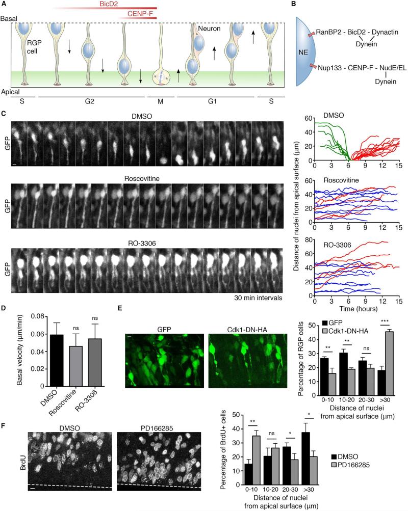
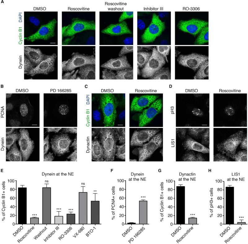
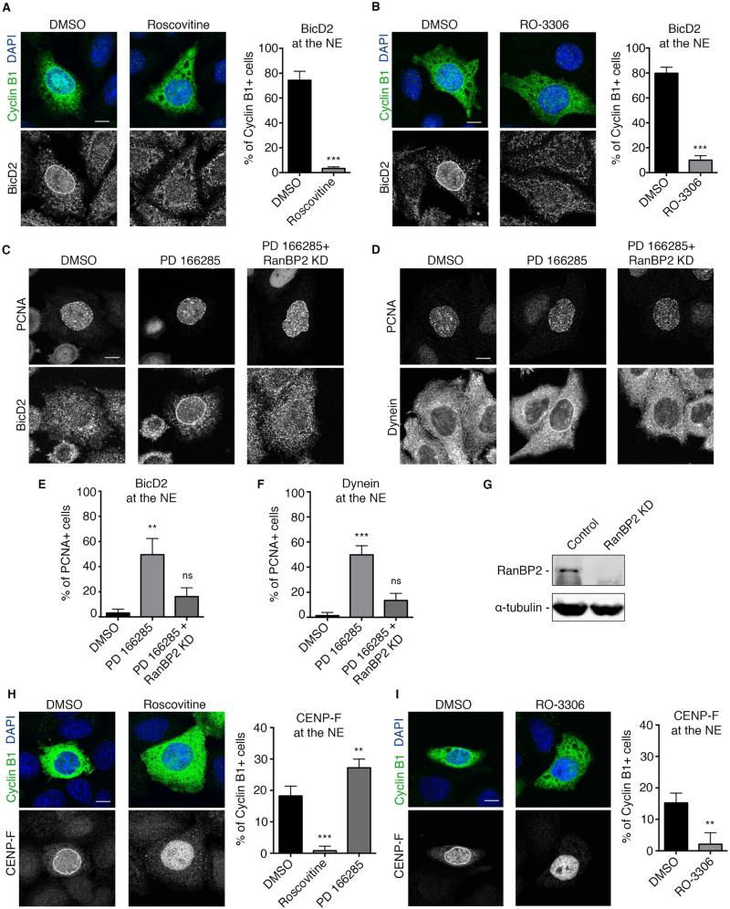
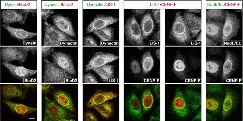
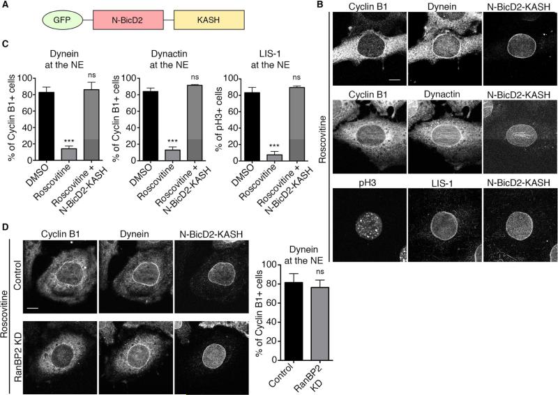
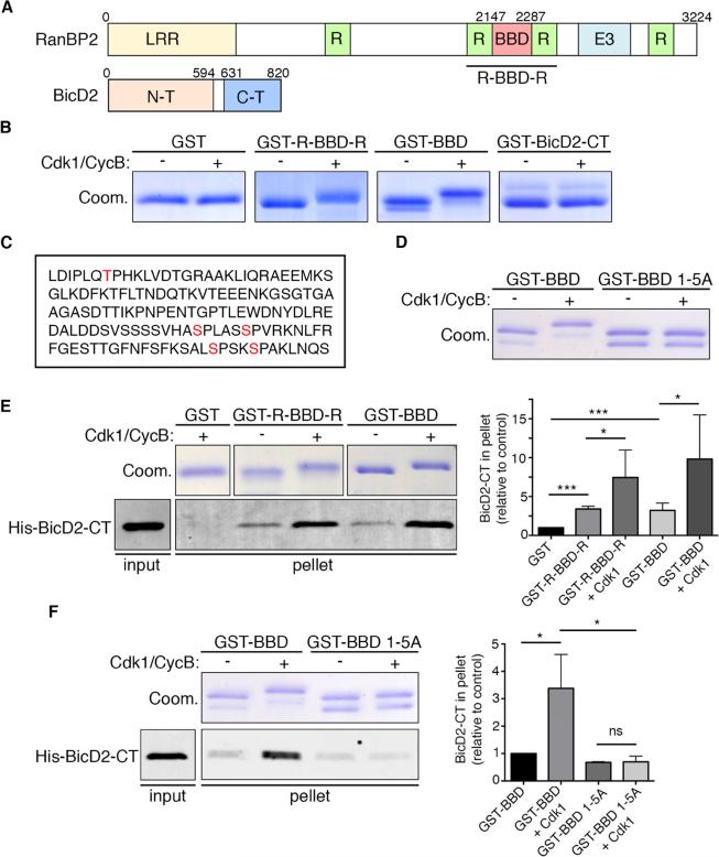
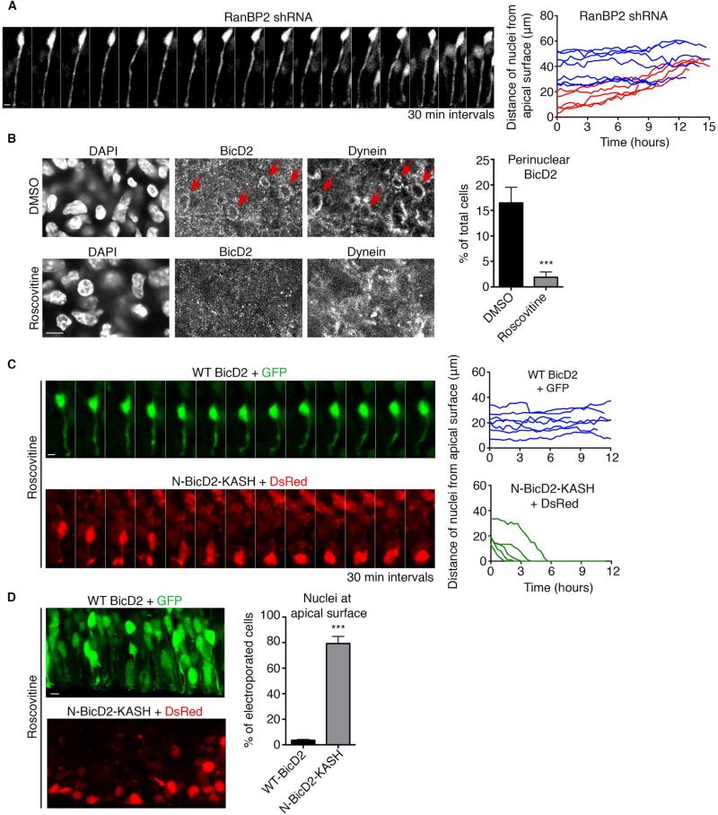
Dynein recruitment to the nuclear envelope is required for pre-mitotic nucleus-centrosome interactions in nonneuronal cells and for apical nuclear migration in neural stem cells. In each case, dynein is recruited to the nuclear envelope (NE) specifically during G2 via two nuclear pore-mediated mechanisms involving RanBP2-BicD2 and Nup133-CENP-F. The mechanisms responsible for cell-cycle control of this behavior are unknown. We now find that Cdk1 serves as a direct master controller for NE dynein recruitment in neural stem cells and HeLa cells. Cdk1 phosphorylates conserved sites within RanBP2 and activates BicD2 binding and early dynein recruitment. Late recruitment is triggered by a Cdk1-induced export of CENP-F from the nucleus. Forced NE targeting of BicD2 overrides Cdk1 inhibition, fully rescuing dynein recruitment and nuclear migration in neural stem cells. These results reveal how NE dynein recruitment is cell-cycle regulated and identify the trigger mechanism for apical nuclear migration in the brain.


Figures

Figure 1
Figure 1. Requirement for Cdk1 in apical nuclear migration in RGP cells
(A) Schematic representation of interkinetic nuclear migration (INM) in RGP cells (from S-phase to S-phase to correspond to time-lapse imaging). Following S-phase, the G2 nucleus moves to the apical (ventricular) surface, driven by NE-associated cytoplasmic dynein. (B) Dynein is recruited to the NE through an early G2 pathway anchored by the nucleoporin RanBP2, responsible for long-range apical nuclear migration; and a late G2 pathway anchored by the nucleoporin Nup133, responsible for pre-mitotic nuclear transport to the ventricular surface of the brain. All factors are expressed throughout the cell cycle except CENP-F, which is absent in G1 and rises during S and G2 phases. (C) Live imaging of GFP-expressing RGP cells in embryonic rat brain slices 3 days after in utero electroporation at E16. The slices were treated with vehicle (DMSO), 55 μM Roscovitine, or 100 μM RO-3306 and imaged for 16 hours (see methods). DMSO-treated cells showed typical INM behavior. Roscovitine and RO-3306 each blocked apical nuclear migration. Right: Representative tracks of individual nuclei for each condition are presented. Green tracks indicate apically migrating nuclei, red tracks basally migrating nuclei, and blue tracks non-migrating nuclei. (D) Basal nuclear migration velocity showed a small, but insignificant change in response to Roscovitine and RO-3306 treatment. (E) Effect of Cdk1 dominant negative on distribution of RGP cells nuclei. Brains were electroporated at E16 with GFP or with Cdk1-DN-HA and fixed and imaged at E18. Measurement of the distance between the bottom of the nucleus and the apical surface shows strong accumulation of nuclei away from the apical surface in cells expressing Cdk1-DN-HA (>30 μm from the ventricle). (F) Effect of Wee1/Myt1 inhibitor PD166285 on distribution of BrdU+ cells, two hours after BrdU pulse. Live brain slices were incubated with BrdU for 15 minutes, washed, and subsequently treated for 2 hours with 1 μM PD166285 or DMSO. The percentage of BrdU+ nuclei that reached the apical region (0-10 μm from the ventricle) strongly increased in PD166285-treated brain slices, compared to DMSO control. For each experiment, at least three independent brains were imaged. Error bars indicate SD; *p<0.05; **p<0.01; ***p<0.001; ns = not significant, based on a Student's t-test. Scale bar, 5 μm. See also Figure S1 and Movies S1, S2, S3, S4 and S5.
Figure 2
Figure 2. Requirement for Cdk1 in HeLa cell nuclear envelope (NE) dynein recruitment
(A) Effect of Cdk1 inhibitors on NE dynein (anti-IC) in G2 (cyclin B1-positive) HeLa cells. Roscovitine (55 μM, 30 min) markedly reduced cytoplasmic dynein staining at the NE, an effect reversed by 30 minutes of Roscovitine washout. Incubation with other Cdk1 inhibitors: inhibitor III (0.9 μM) or RO-3306 (28 μM), for 30 minutes, also blocked dynein accumulation at the NE. (B) Effect of Wee1/Myt1 inhibitor PD166285 on dynein recruitment to the NE. HeLa cells were immunostained for dynein (IC) and the S phase marker PCNA (which forms intranuclear foci at this stage). Exposure to PD166285 (0.5 μM, 30 min) strongly increased the fraction of S phase cells exhibiting NE dynein. (C) Roscovitine (55 μM, 30 min) strongly inhibited NE dynactin (anti-p150) staining in Cyclin B1-positive HeLa cells and (D) NE LIS1 staining in phospho-Histone 3 (pH3)-positive HeLa cells (For antibody compatibility, phospho-histone H3 (pH3) was used as a marker for G2/prophase cells, see methods). (E) Quantification of the effects of Cdk1 inhibitors Roscovitine, Inhibitor III, and RO-3306; Aurora A inhibitor VX-680 (0.3 μM); and Plk1 inhibitor BTO-1 (22 μM) on dynein recruitment to the NE of cyclin B1+ cells. (F) Quantification of the effect of Wee1/Myt1 inhibitor PD166285 on dynein recruitment to the NE of S-phase (PCNA+) cells. (G) Quantification of the effect of Cdk1 inhibitor Roscovitine on dynactin recruitment to the NE of cyclin B1+ cells. (H) Quantification of the effect of Cdk1 inhibitor Roscovitine on LIS1 recruitment to the NE of pH3+ cells. Each experiment was reproduced three independent times (over 50 cells per condition and per experiment were counted). Error bars indicate SD; **p<0.01; ***p<0.001; ns = not significant, based on a Student's t-test. Scale bar, 10 μm.
Figure 3
Figure 3. Requirement for Cdk1 in both early and late dynein NE recruitment pathways
(A) Roscovitine (55 μM, 30 min) strongly inhibited BicD2 recruitment to the NE of Cyclin B1+ cells. (B) RO-3306 (28 μM, 30 min) also strongly impaired BicD2 recruitment to the NE of Cyclin B1+ cells. (C) The Wee1/Myt1 inhibitor PD166285 (0.5 μM, 30 min) induced premature BicD2 accumulation at the S-phase NE marked by PCNA. siRNA-mediated knockdown of RanBP2 inhibited this premature BicD2 recruitment in PCNA+ cells. (D) siRNA-mediated knockdown of RanBP2 also inhibited the premature recruitment of dynein to the NE of S phase cells (marked by PCNA). (E and F) Quantification of the effect of Wee1/Myt1 inhibitor PD166285 and RanBP2 knockdown on (E) BicD2 and (F) dynein recruitment to the nuclear envelope of S phase cells (marked by PCNA). (G) Western Blot analysis of RanBP2 levels in HeLa cells reveals strong knockdown 3 days post-transfection with RanBP2 siRNA. (H) Effect of Roscovitine and PD166285 on CENP-F recruitment to the NE of cyclin B1+ cells. NE CENP-F decreased from 16.75 ± 1.7 % in DMSO-treated cells to 0.82 ± 1.4 % in Roscovitine-treated cells but increased to 27.2 ± 1.6% in PD166285 treated cells. Roscovitine treatment caused CENP-F to remain inside the nucleus. (I) RO-3306 (28 μM, 30 min) also strongly impaired CENP-F recruitment to the NE of Cyclin B1+ cells, which remained inside the nucleus. Each experiment was reproduced three independent times (over 50 cells per condition and per experiment were counted). Error bars indicate SD; **p<0.01, ***p<0.001, ns = not significant, based on a Student's t-test. Scale bar, 10 μm.
Figure 4
Figure 4. Colocalization analysis between dynein and dynein regulatory factors at the NE
Stainings for dynein and known dynein partners at the NE. Factors were stained two by two and colocalization was assessed. Dynein, dynactin, BicD2 and LIS1 always colocalize to the nuclear envelope, suggesting that they are all part of the early dynein recruitment pathway. LIS1 and CENP-F only colocalize in 23.6% (± 2.4%) of the case, while in 76.4% (± 2.4%) of LIS1-positive cells, CENP-F is still intranuclear. CENP-F and NudE/EL always colocalize at the NE, suggesting that they are both specific to the late dynein recruitment pathway.
Figure 5
Figure 5. Forced recruitment of BicD2 to the NE rescues dynein localization in Cdk1-inhibited cells
(A) Schematic representation of N-BicD2-KASH fusion protein (Splinter et al., 2010). The C-terminal RanBP2-binding domain has been replaced by the KASH nuclear envelope targeting motif from Nesprin 3 (yellow). (B) Effect of NE-targeted BicD2 on NE envelope recruitment of dynein and its cofactors in Cdk1-inhibited HeLa cells. Cells were transfected with N-BicD2-KASH, treated with Roscovitine (55 μM, 30 min), and stained for dynein, dynactin, and LIS1. N-BicD2-KASH decorates the NE and restores dynein, dynactin, and LIS1 localization to the NE of cyclin B1+ cells in the first two cases, and of pH3+ cells in the case of LIS1. (C) Quantification of the effect of N-BicD2-KASH expression on dynein, dynactin, and LIS1 localization to the NE in Roscovitine treated cells. (D) RanBP2 knockdown did not affect N-BicD2-KASH-mediated recruitment of dynein/dynactin to the NE of Cyclin B1+ cells. Each experiment was reproduced three independent times (over 50 cells per condition and per experiment were counted). Error bars indicate SD; ***p<0.001; ns = not significant, based on a Student's t-test. Scale bar, 10 μm. See also Figures S2 and S3.
Figure 6
Figure 6. Effect of Cdk1 phosphorylation on RanBP2-BicD2 interaction
(A) Schematic representation of RanBP2 and BicD2. RanBP2 is a large, multi-functional nucleoporin containing a leucine-rich region (LRR), four Ran-binding domains (R), an E3 Sumo ligase domain (E3), and a BicD2-bindng domain (BBD). BicD2 contains substantial regions of α-helical coiled-coil and binds the dynein and dynactin complexes through its N-terminal region and other factors, including RanBP2, through its C-terminal cargo-binding domain (Liu et al., 2013). (B) In vitro assay for Cdk1 phosphorylation of GST-tagged RanBP2 BBD, RanBP2 R-BBD-R, and BicD2-CT. Both of the BicD2-binding RanBP2 fragments displayed a shift in migration on Coomassie-stained electrophoretic gels (Coom.) upon exposure to in vitro purified recombinant Cdk1 and Cyclin B in the presence of ATP. GST-BicD2-CT and GST alone showed no such effect. (C) RanBP2 BBD sequence (amino acids 2147 to 2287) showing the five Cdk1 phosphorylation sites (red) identified on the basis of the known Cdk1 consensus motif and directly demonstrated to be phosphorylated by Cdk1 in vitro using mass spectrometry. (D) In vitro Cdk1 phosphorylation of GST-BBD WT and mutated to alanine in all five identified Cdk1 phosphorylation sites (GST-BBD 1-5A). The electrophoretic gel shift observed for the WT fragment was abolished in the BBD 1-5A fragment. (E) GST-pull down assay with indicated RanBP2 fusions and purified His-BicD2-CT in the presence or absence of purified Cdk1/cyclin B. GST fragments were visualized by Coomassie Blue staining, and His-BicD2-CT was detected by Western blotting with anti-His tag antibody. 10% of the input and 50% of the bound fractions were loaded on the gel. Right: Quantification of BicD2-CT bound fraction relative to amount bound to GST (n=5 independent experiments). Phosphorylation of the RanBP2 fusions by Cdk1 dramatically increased binding to BicD2-CT. (F) GST-pull down assay with indicated RanBP2 fusions and purified His-BicD2-CT in the presence or absence of purified Cdk1 + cyclin B. 10% of the input and 50% of the bound fractions were loaded on gel. Right: Quantification BicD2-CT bound fraction relative to amount bound to GST-BBD (n=4 independent experiments). Pull down with GST-BBD 1-5A shows loss of Cdk1-dependant affinity increase for BicD2-CT. Error bars indicate SD; *p<0.05; ***p<0.001; ns = not significant, based on a Student's t-test. See also Figure S4.
Figure 7
Figure 7. Role of RanBP2-regulated recruitment of BicD2 to the NE of RGP cells
(A) Live imaging of RanBP2 shRNA-expressing RGP cells in embryonic rat brain slices 4 days after in utero electroporation at E16. The nuclei of electroporated cells were unable to undergo apical nuclear migration. Right: Representative tracings of nuclei. Red tracks indicate basally migrating nuclei and blue tracks non-migrating nuclei. Scale bar, 5 μm. (B) NE BicD2 and dynein labeling within the ventricular zone (VZ) of E19 rat brain sections. Top: NE BicD2 and dynein staining was observed in a subset of cells in control DMSO-treated brain slices (red arrows). Bottom: NE BicD2 and dynein staining was absent from the NE of RGP cells in brain slices treated with Roscovitine (55 μM, 60 min prior to fixation). Right: Quantification of the percentage of total cells with NE BicD2 staining. Only nuclei located within 40 μm from the ventricular surface were evaluated. (C) Live imaging of RGP cell expressing wild type BicD2 + GFP or N-BicD2-KASH + DsRed and treated with Roscovitine (55 μM). Brains were subjected to in utero electroporation at E18 and sliced and put into culture at E19. Top: Wild type BicD2 expression does not restore apical nuclear migration in the presence of Roscovitine. Bottom: N-BicD2-KASH restores apical nuclear migration in the presence of Roscovitine. Green tracks indicate apically migrating nuclei and blue tracks non-migrating nuclei. (D) Brains were subjected to electroporation at E19 and directly sliced and put into culture in the presence of Roscovitine (55 μM) for 24 hours, before fixation. Top: Nuclei of RGP cells expressing wild type BicD2 + GFP do not reach the apical surface in the presence of Roscovitine. Bottom: A high proportion of nuclei are at the apical surface in RGP cells expressing N-BicD2-KASH + DsRed. Right: Quantification of the percentage of nuclei reaching the apical surface. For each experiment, at least three independent brains were imaged. Error bars indicate SD; ***p<0.001, based on a Student's t-test. Scale bar, 5 μm. See also Figure S5 and Movies S6, S7, S8 and S9.

References

    1. Bader JR, Kasuboski JM, Winding M, Vaughan PS, Hinchcliffe EH, Vaughan KT. Polo-like kinase1 is required for recruitment of dynein to kinetochores during mitosis. Journal of Biological Chemistry. 2011;286:20769–20777. - PMC - PubMed
    1. Beaudouin J, Gerlich D, Daigle N, Eils R, Ellenberg J. Nuclear envelope breakdown proceeds by microtubule-induced tearing of the lamina. Cell. 2002;108:83–96. - PubMed
    1. Bolhy S, Bouhlel I, Dultz E, Nayak T, Zuccolo M, Gatti X, Vallee R, Ellenberg J, Doye V. A Nup133-dependent NPC-anchored network tethers centrosomes to the nuclear envelope in prophase. J Cell Biol. 2011;192:855–871. - PMC - PubMed
    1. Cai Y, Singh BB, Aslanukov A, Zhao H, Ferreira PA. The docking of kinesins, KIF5B and KIF5C, to Ran-binding protein 2 (RanBP2) is mediated via a novel RanBP2 domain. J Biol Chem. 2001;276:41594–41602. - PubMed
    1. Deibler RW, Kirschner MW. Quantitative reconstitution of mitotic CDK1 activation in somatic cell extracts. Mol Cell. 2010;37:753–767. - PMC - PubMed

Publication types

MeSH terms

LinkOut - more resources