. 2015 Jun 15;25(12):1654-60.
doi: 10.1016/j.cub.2015.04.058. Epub 2015 Jun 4.

Replication fork progression during re-replication requires the DNA damage checkpoint and double-strand break repair

Affiliations

Replication fork progression during re-replication requires the DNA damage checkpoint and double-strand break repair

Jessica L Alexander et al. Curr Biol. .

Abstract

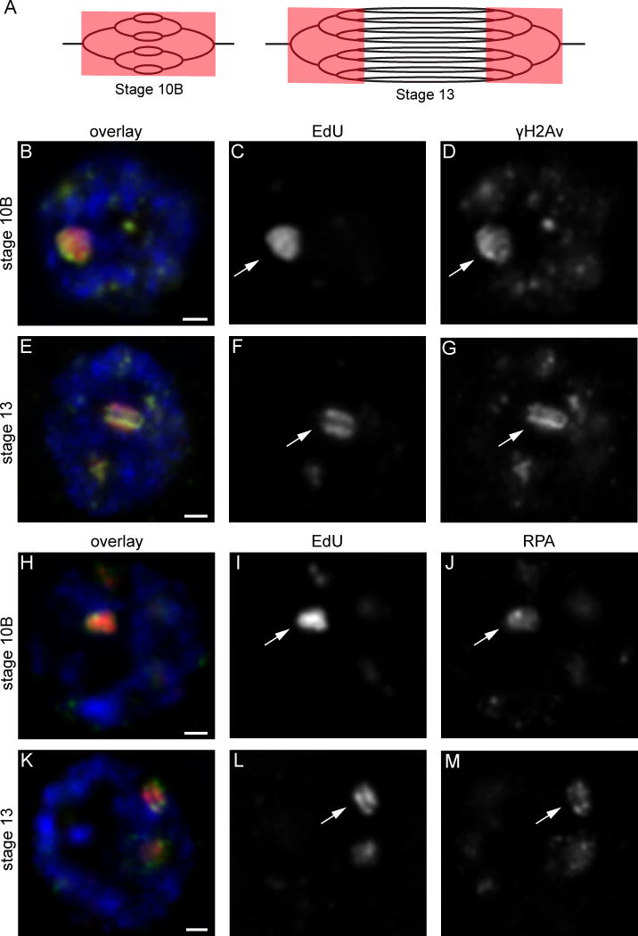
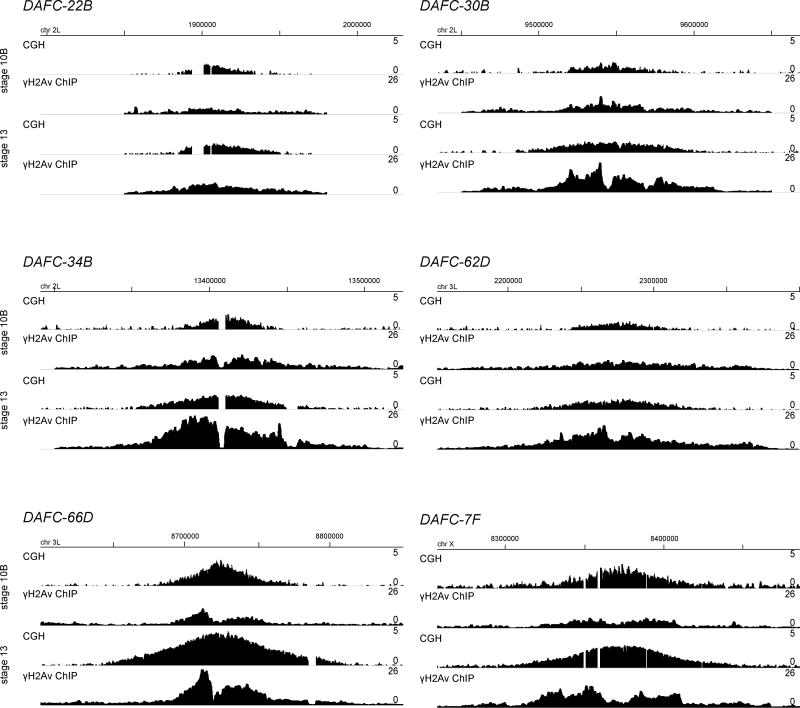
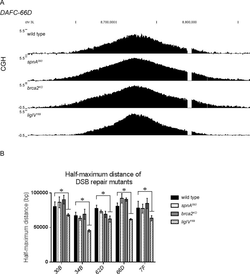
Replication origins are under tight regulation to ensure activation occurs only once per cell cycle [1, 2]. Origin re-firing in a single S phase leads to the generation of DNA double-strand breaks (DSBs) and activation of the DNA damage checkpoint [2-7]. If the checkpoint is blocked, cells enter mitosis with partially re-replicated DNA that generates chromosome breaks and fusions [5]. These types of chromosomal aberrations are common in numerous human cancers, suggesting that re-replication events contribute to cancer progression. It was proposed that fork instability and DSBs formed during re-replication are the result of head-to-tail collisions and collapse of adjacent replication forks [3]. However, previously studied systems lack the resolution to determine whether the observed DSBs are generated at sites of fork collisions. Here, we utilize the Drosophila ovarian follicle cells, which exhibit re-replication under precise developmental control [8-10], to model the consequences of re-replication at actively elongating forks. Re-replication occurs from specific replication origins at six genomic loci, termed Drosophila amplicons in follicle cells (DAFCs) [10-12]. Precise developmental timing of DAFC origin firing permits identification of forks at defined points after origin initiation [13, 14]. Here, we show that DAFC re-replication causes fork instability and generates DSBs at sites of potential fork collisions. Immunofluorescence and ChIP-seq demonstrate the DSB marker γH2Av is enriched at elongating forks. Fork progression is reduced in the absence of DNA damage checkpoint components and nonhomologous end-joining (NHEJ), but not homologous recombination. NHEJ appears to continually repair forks during re-replication to maintain elongation.


Figures

Figure 1
Figure 1. Markers of DNA damage and replication fork stress co-localize with sites of re-replication
(A) The onion skin model of amplification. EdU is drawn in red overlaying sites of actively replicating DNA. EdU labeling during origin initiation and fork progression in stage 10B results in incorporation throughout the amplicons (left). In stage 13 when forks continue to progress without further origin firing events, EdU incorporation gives rise to the double-bar structure (right). (B–G) Immunofluorescence images of stage 10B (B–D) and 13 (E–G) follicle cell nuclei reveal the double-strand break marker γH2Av (D, G) co-localizes with EdU (C, F). As forks progress in stage 13 and EdU incorporation forms the double-bar structure (F), the γH2Av signal also resolves into to double-bars (G). This co-localization pattern was present in every follicle cell nucleus of every egg chamber observed (53 stage 10Bs and 49 stage 13s). (B, E) Merged image with EdU is shown in red, γH2Av in green, DAPI in blue. Each image is a single plane of nucleus. The prominent EdU focus corresponds to DAFC-66D (arrows). Scale bars, 1μm. (H–M) RPA immunofluorescence reveals direct overlap with EdU in stage 10B (H–J) and 13 (K–M) follicle cells. RPA follows the pattern of fork progression highlighted by EdU, resolving into a double-bar structure in stage 13 (M). This co-localization pattern was present in every follicle cell nucleus of every egg chamber observed (51 stage 10Bs and 60 stage 13s). (H, K) Merged image with EdU is shown in red, RPA in green, DAPI in blue. Each image is a single plane of a follicle cell nucleus. The prominent EdU focus corresponds to DAFC-66D (arrows). Scale bars, 1 μm.
Figure 2
Figure 2. γH2Av enrichment at the DAFCs during re-replication stages
CGH and γH2Av ChIP-seq from OrR stage 10B and 13 follicle cells at each of the six DAFCs. Chromosomal position is indicated above each panel. CGH profiles are the log2 ratio (0–5) of egg chamber to embryonic DNA (first and third lines). ChIP-seq enrichment is the RPM of ChIP/input (0–26) for 1kb windows sliding every 100bp, and is the geometric mean of two biological replicates (second and fourth lines). Genomic coordinates are displayed above.
Figure 3
Figure 3. Fork progression is reduced in the absence of DDR components
(A) Blocked fork progression causes adjacent forks to pile up, resulting in close spacing as demonstrated by the replication forks highlighted in red (top). This is reflected in the CGH gradient by a sharp decrease in copy number. An example of one such region is highlighted in red on the wild-type DAFC-66D gradient (bottom). (B) CGH of DAFC-66D from DDR mutants reveals impaired replication fork progression. DNA from stage 13 egg chambers was competitively hybridized with diploid embryonic DNA to microarrays with approximately one probe every 125bp. Chromosomal position is plotted on the x-axis, the log2 ratio of stage 13 DNA to embryonic DNA is plotted on the y-axis. In all mutants shown, the amplification gradient exhibits a rapid decrease in copy number compared to the wild type (top). (C) The half-maximum distance was calculated in the wild-type and mutant backgrounds for each DAFC. Each half-maximum value is the average of three biological replicates. Significance measured by the Dunnett test for multiple comparisons, asterisks indicate p<0.05 and n.s. indicates not significant. (D) The level of amplification was measured at the DAFC-66D origin of replication in each DSB signaling and repair mutant by quantitative real-time PCR. The copy number in stages 10B and 13 egg chambers is relative to the nonamplified rosy locus. Error bars are standard error of three replicates. None of the mutants were significantly different from the wild type as measured by the Dunnett test for multiple comparisons.
Figure 4
Figure 4. LigIV is utilized for DSB repair during re-replication
(A) CGH of DAFC-66D reveals impaired replication fork progression in the ligIV169, but not the spnA093 or brca2KO mutants. DNA from stage 13 egg chambers was competitively hybridized with diploid embryonic DNA to microarrays with approximately one probe every 125bp. Chromosomal position is plotted on the x-axis, the log2 ratio of stage 13 DNA to embryonic DNA is plotted on the y-axis. (B) The half-maximum distance was calculated in the wild-type and mutant backgrounds for each DAFC. Each half-maximum value is the average of three biological replicates. Significance measured by the Dunnett test for multiple comparisons, asterisks indicate p<0.05. The spnA093 and brca2KO mutants are not significantly different from wild type.

References

    1. Bell SP, Dutta A. DNA replication in eukaryotic cells. Annu Rev Biochem. 2002;71:333–374. - PubMed
    1. Abbas T, Keaton MA, Dutta A. Genomic instability in cancer. Cold Spring Harb Perspect Biol. 2013;5:a012914. - PMC - PubMed
    1. Davidson IF, Anatoily L, Blow JJ. Deregulated replication licensing causes DNA fragmentation consistent with head-to-tail fork collision. Mol Cell. 2006;24:433–443. - PMC - PubMed
    1. Green BM, Li JJ. Loss of rereplication control in Saccharomyces cerevisiae results in extensive DNA damage. Mol Biol Cell. 2005;16:421–432. - PMC - PubMed
    1. Melixetian M, Ballabeni A, Masiero L, Gasparini P, Zamponi R, Bartek J, Lukas J, Helin K. Loss of Geminin induces rereplication in the presence of functional p53. J Cell Biol. 2004;165:473–482. - PMC - PubMed

Publication types