. 2015 Jun 23;11(11):1760-71.
doi: 10.1016/j.celrep.2015.05.021. Epub 2015 Jun 4.

Quaternary Structure Defines a Large Class of Amyloid-β Oligomers Neutralized by Sequestration

Affiliations

Quaternary Structure Defines a Large Class of Amyloid-β Oligomers Neutralized by Sequestration

Peng Liu et al. Cell Rep. .

Abstract

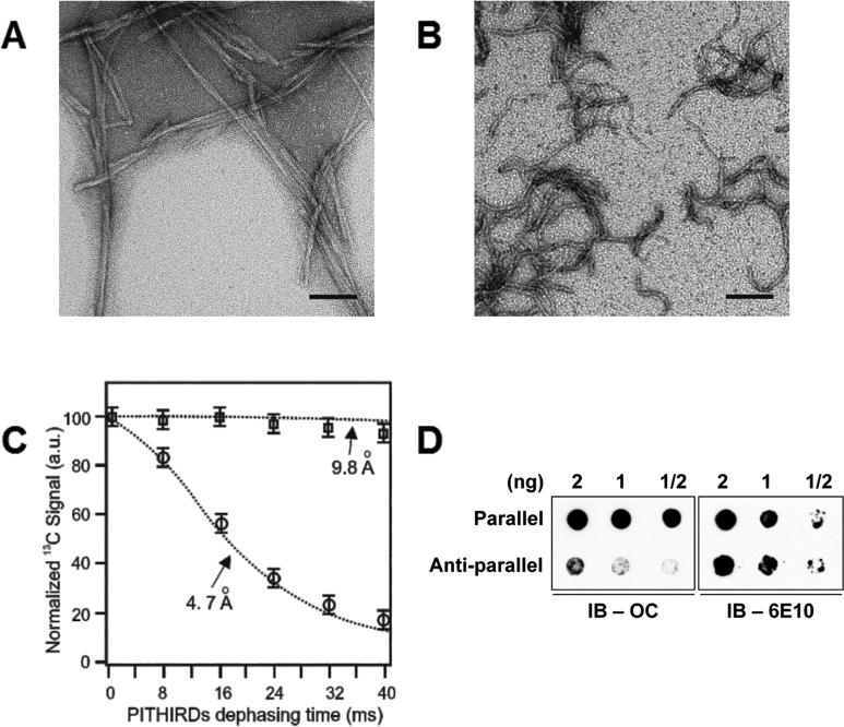
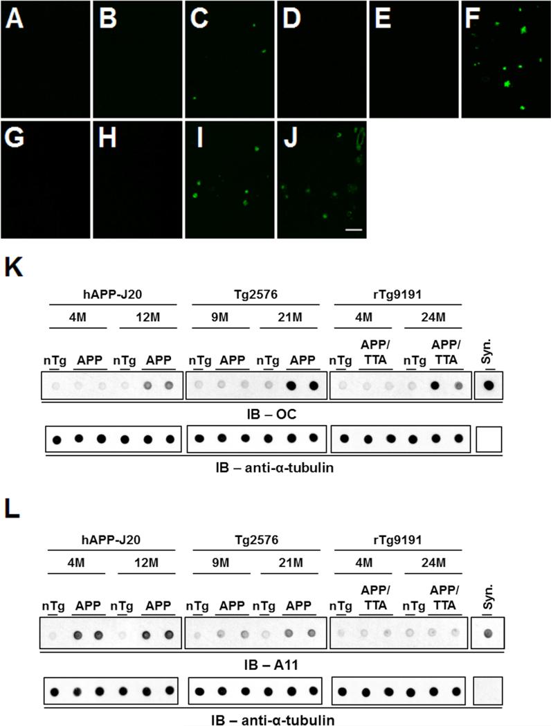
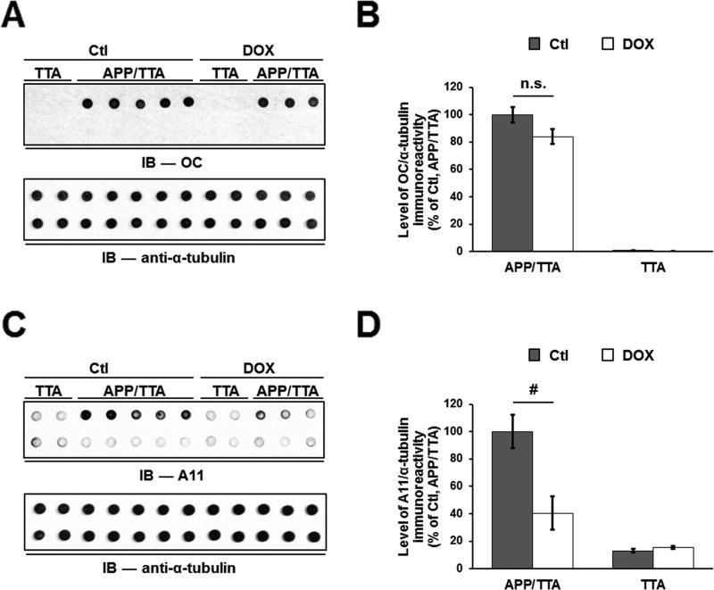
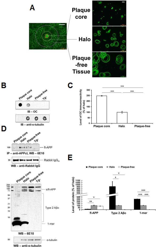
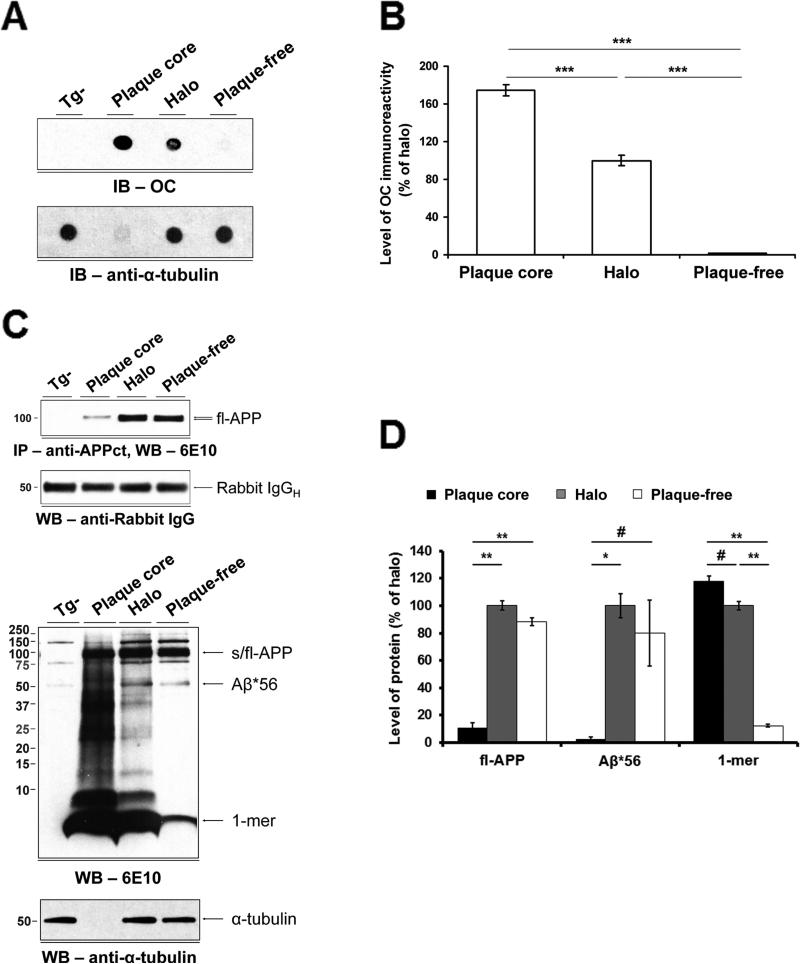
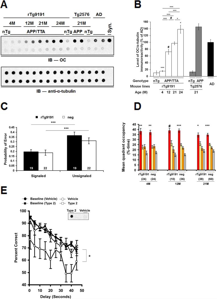
The accumulation of amyloid-β (Aβ) as amyloid fibrils and toxic oligomers is an important step in the development of Alzheimer's disease (AD). However, there are numerous potentially toxic oligomers and little is known about their neurological effects when generated in the living brain. Here we show that Aβ oligomers can be assigned to one of at least two classes (type 1 and type 2) based on their temporal, spatial, and structural relationships to amyloid fibrils. The type 2 oligomers are related to amyloid fibrils and represent the majority of oligomers generated in vivo, but they remain confined to the vicinity of amyloid plaques and do not impair cognition at levels relevant to AD. Type 1 oligomers are unrelated to amyloid fibrils and may have greater potential to cause global neural dysfunction in AD because they are dispersed. These results refine our understanding of the pathogenicity of Aβ oligomers in vivo.


Figures

Figure 1
Figure 1. OC antibodies recognize in-register parallel β-sheet structures
Transmission electron micrographs show D23N_Aβ40 fibrils with (A) in-register parallel β-sheet structure and (B) anti-parallel structure. (C) 13C-PITHIRDs-CT decay curves for parallel and anti-parallel fibrils with 13C labeling at Ala21-13C’. Theoretical decay curves with 4.7 and 9.8 angstrom 13C-13C distances are shown as dotted lines. Experimental data for the parallel and anti-parallel fibrils are indicated by circles and squares, respectively. The error bars were determined from the experimental spectral noise. (D) Left panel, dot blot shows that OC antibodies selectively detect D23N_Aβ40 fibrils with in-register parallel β-sheet structure. Right panel, blot shown at left was stripped and re-probed with monoclonal antibody 6E10 to confirm that ~equal amounts of parallel and anti-parallel fibrils were loaded. Scale bars (A,B): 100 nm. See also Figure S1.
Figure 2
Figure 2. Age-dependent appearance of A11- and OC-immunoreactive Aβo
(A-J) Brain sections stained with Thioflavin S to reveal dense-core plaques in cerebral cortex. A-C: hAPP-J20 (A, non-transgenic, 4M; B, hAPP-J20, 4M; C, hAPP-J20, 12M); D-F: Tg2576 (D, non-transgenic, 9M; E, Tg2576, 9M; F, Tg2576, 21M); G-I: rTg9191 (G, non-transgenic, 4M; H, rTg9191, 4M; I, rTg9191, 24M); J: AD brain. Scale bar in (J), 100 μm, applies to (A-J). (K) OC-reactive aggregates are seen after the appearance of dense-core plaques. Upper panels, dot blots showing protein aggregates detected by polyclonal OC antibodies in water-soluble brain extracts from hAPP-J20, Tg2576 and rTg9191 mice prior to (hAPP-J20, 4M; Tg2576, 9M; rTg9191, 4M) and after (hAPP-J20, 12M; Tg2576, 21M; rTg9191, 24M) the appearance of dense-core plaques. Extracts from non-transgenic littermates (nTg) are shown for comparison. Syn, synthetic soluble Aβ aggregates with in-register parallel β-sheets (2 ng) were used as a positive control. Lower panels, α-tubulin served as the loading control. (L) A11-reactive aggregates are seen prior to the appearance of dense-core plaques. Upper panels, dot blots, prepared as in (K), but probed with A11 antibodies. Syn, synthetic Aβ40 oligomers prepared as in (Kayed et al., 2003). Lower panels, α-tubulin loading control. See also Figures S2 and S7.
Figure 3
Figure 3. Suppression of transgenic APP selectively lowers A11-immunoreactive Aβo in TetO-APPSweInd mice
(A) Upper panel, protein aggregates detected by OC antibodies in aqueous brain extracts prepared from 8.5-month-old TetO-APPSweInd mice harboring both activator and responder transgenes (APP/TTA) or only the activator transgene (TTA). DOX, mice administered doxycycline to suppress APP expression for the 5 weeks immediately preceding sacrifice; Ctl, untreated mice. Upper lane, brain extracts; lower lane, brain extracts immunodepleted of Aβ. Lower panel, α-tubulin loading control. (B) Quantification of OC immunoreactivity. There is no difference between levels of OC-immunoreactivity, normalized to α-tubulin levels, in extracts from control (Ctl) and DOX-treated mice. (C) Upper panel, protein aggregates detected by A11 in the extracts described in (A). Upper lane, brain extracts; lower lane, brain extracts immunodepleted of Aβ. Lower panel, α-tubulin loading control. (D) Quantification of A11 immunoreactivity. Suppression of transgenic APP expression in TetO-APPSweInd mice resulted in a ~60% reduction in the levels of A11-reactive oligomers. n.s. = not significant, # p < 0.05, twoway ANOVA followed by Fisher's post-hoc analysis. See also Figure S7.
Figure 4
Figure 4. Type 2 Aβo are confined to dense-core plaques in rTg9191 mice
(A) Photomicrographs of microdissected fractions. White scale bars: 100 μm. (B) Upper panel, dot blot, probed with OC antibodies, illustrates the topographical distribution of native Type 2 Aβo; lower panel, above blot stripped and re-probed with anti-α-tubulin, showing that equal amounts of protein were loaded for the halo and plaque-free regions. (C) Quantification of dot blots. OC-reactive aggregates are confined to the dense-core and halo regions. (D) Immunoblotting under denaturing conditions confirms that Type 2 Aβo are restricted to the dense-core and halo regions. Upper two panels, Western blot showing full-length APP (fl-APP) in detergent-soluble extracts of laser microdissected fractions. Fl-APP was immunoprecipitated with a polyclonal antibody directed against a C-terminal epitope in APP (anti-APPct) and detected using 6E10 (top). Blot stripped and re-probed with an antibody directed against rabbit immununoglobulin (IgG) showing that ~equal amounts of capture antibody were immunoprecipitated (bottom). Lower two panels, Western blot probed with monoclonal antibody 6E10 (top). Blot stripped and re-probed with anti-α-tubulin (bottom). (E) Quantification of Western blots. Type 2 Aβo and monomers reside in the dense-core and halo regions, while fl-APP is primarily found in halo and plaque-free regions. * p < 0.01, ** p < 0.001, *** p < 0.0001, one-way ANOVA followed by Fisher's post hoc analysis. Abbreviations: 1-mer, Aβ monomer; s/fl-APP, soluble and full-length amyloid precursor protein; Tg-, nontransgenic littermates of rTg9191 mice; IgGH, immunoglobulin heavy chain. See also Figures S3, S4 and S7.
Figure 5
Figure 5. Type 2 Aβo and Aβ*56 have different spatial distributions in the brains of Tg2576 mice
(A) Upper panel, dot blot probed with OC antibodies shows the topographical distributions of Type 2 Aβo; lower panel, α-tubulin blot shows that equal amounts of protein were loaded for the halo and plaque-free regions. (B) Quantification of dot blots. Type 2 Aβo overwhelmingly reside in the dense-core and halo regions. (C) Upper two panels, Western blot showing fl-APP in detergent-soluble extracts of laser microdissected fractions (top). Blot stripped and re-probed with anti-rabbit IgG antibody showing that ~equal amounts of capture antibody were immunoprecipitated (bottom). Lower two panels, Western blot probed with 6E10 antibody shows the topographical distributions of APP, Aβ*56 and monomers (1-mer) (top). Alpha-tubulin blot shows that equivalent amounts of protein were loaded in the halo and plaque-free lanes (bottom). (D) Quantification of Western blots. Aβ*56 and fl-APP are primarily found in halo and plaque-free regions. In contrast, Aβ monomers overwhelmingly reside in the dense-core and halo regions. # p < 0.05, * p < 0.01, ** p < 0.001, *** p < 0.0001, one-way ANOVA followed by Fisher's post-hoc analysis. Tg-, non-transgenic littermates of Tg2576; other abbreviations as in Figure 4. See also Figures S3, S4 and S7.
Figure 6
Figure 6. Type 2 Aβo do not disrupt cognition when located in situ in rTg9191 brains, but do impair cognition when dispersed
(A-D) rTg9191 mice producing levels of Type 2 Aβo comparable to those of AD patients have intact cognition. (A) Upper panel, Dot blot showing the relative levels of soluble OC-reactive aggregates in the brains of rTg9191 mice, non-transgenic littermates (nTg) and AD patients. Ages of mice are shown above the blot. Upper lane, water-soluble brain extracts and synthetic aggregates; lower lane, samples immunodepleted of Aβ. Syn, synthetic soluble Aβ aggregates with in-register parallel β-sheets. Lower panel, α-tubulin served as the loading control for both the untreated (upper lane) and Aβ-immunodepleted extracts (lower lane). (B) Quantification of dot blots. Levels of OC-reactive aggregates in the brains of rTg9191 mice increase with age (# p < 0.05, * p < 0.01, *** p < 0.0001, one-way ANOVA followed by Fisher's post hoc analysis). (C) Cognitive performance in 23-month-old APP- positive (rTg9191) and negative (neg) rTg9191 mice do not differ in the Signaled and Unsignaled components of the fixed consecutive number (FCN-4) test. The probability of a given trial producing an error in the Signaled component is significantly lower than in the Unsignaled component, indicating intact motor and visual function (*** p < 0.0001, paired t-test). (D) Spatial reference memory in rTg9191 mice does not differ significantly from that of APP-neg littermates. Young (4M), middle-aged (12M) and old (21M) mice were tested in the water maze; mean %-time in each quadrant (target, red; left of target, yellow; right of target, orange; and opposite the target, grey) is shown. Significant search biases favoring the target quadrant were found in both rTg9191 mice and APP-neg littermates at all three ages. # p < 0.05, * p < 0.01, ** p < 0.001, *** p < 0.0001, percentage time spent in the target compared to that in each of the other three quadrants, repeated measures ANOVA (RMANOVA) followed by Fisher's post hoc analysis. Numerals in (C) and (D) represent the number of mice tested. (E) Water-soluble brain extracts of rTg9191 mice containing Type 2 Aβo disrupt cognition when exogenously administered to rats. Rats (N = 18) previously trained on a delayed non-matched to position task were injected intracerebroventricularly with brain extracts of rTg9191 mice containing Type 2 Aβo or with extracts immunodepleted of Aβ (Vehicle). Type 2 Aβo, but not Vehicle, injections impaired performance on this task (* P < 0.01, RMANOVA compared to baseline performance (mean of three contiguous sessions)). Inset: dot blot showing OC-immunoreactivity in Type 2 Aβo-containing (“Type 2”) and immunodepleted (“Vehicle”) injectates. See also Figure S6 and Table S1.

References

    1. Alzforum Bapineuzumab. 2015a
    1. Alzforum Gantenerumab. 2015b
    1. Benilova I, Karran E, De Strooper B. The toxic Abeta oligomer and Alzheimer's disease: an emperor in need of clothes. Nat Neurosci. 2012;15:349–357. - PubMed
    1. Billings LM, Green KN, McGaugh JL, LaFerla FM. Learning decreases A beta*56 and tau pathology and ameliorates behavioral decline in 3xTg-AD mice. J Neurosci. 2007;27:751–761. - PMC - PubMed
    1. Bolmont T, Haiss F, Eicke D, Radde R, Mathis CA, Klunk WE, Kohsaka S, Jucker M, Calhoun ME. Dynamics of the microglial/amyloid interaction indicate a role in plaque maintenance. J Neurosci. 2008;28:4283–4292. - PMC - PubMed

Publication types

MeSH terms

Substances