. 2015 Jun 16;11(10):1625-37.
doi: 10.1016/j.celrep.2015.05.019. Epub 2015 Jun 4.

ASXL2 Regulates Glucose, Lipid, and Skeletal Homeostasis

Affiliations

ASXL2 Regulates Glucose, Lipid, and Skeletal Homeostasis

Takashi Izawa et al. Cell Rep. .

Abstract

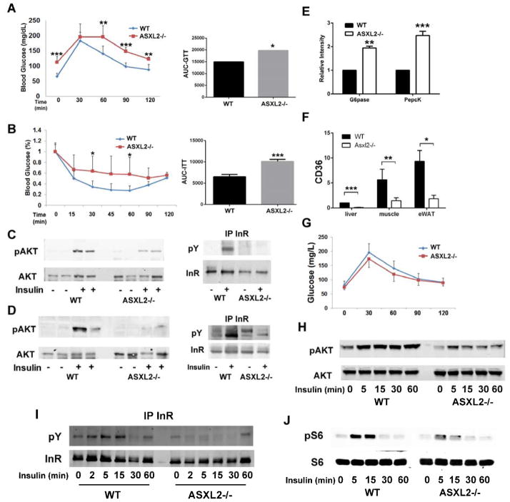
ASXL2 is an ETP family protein that interacts with PPARγ. We find that ASXL2-/- mice are insulin resistant, lipodystrophic, and fail to respond to a high-fat diet. Consistent with genetic variation at the ASXL2 locus and human bone mineral density, ASXL2-/- mice are also severely osteopetrotic because of failed osteoclast differentiation attended by normal bone formation. ASXL2 regulates the osteoclast via two distinct signaling pathways. It induces osteoclast formation in a PPARγ/c-Fos-dependent manner and is required for RANK ligand- and thiazolidinedione-induced bone resorption independent of PGC-1β. ASXL2 also promotes osteoclast mitochondrial biogenesis in a process mediated by PGC-1β but independent of c-Fos. Thus, ASXL2 is a master regulator of skeletal, lipid, and glucose homeostasis.


Figures

Fig 1
Fig 1
ASXL2 deficiency results in impaired osteoclastogenesis. A) WT and ASXL2−/− BMMs were cultured with M-CSF and RANKL for 5 days and osteoclasts were counted. B) Osteoclast differentiation proteins were determined by immunoblot, with time. C) BMMs were cultured on bone with M-CSF and RANKL and medium CTx determined. D) ASXL2−/−BMMs, transduced with ASXL2 or vector, were exposed to M-CSF and RANKL for 5 days. The cells were TRAP stained. Vector-transduced WT cells serve as control. E) Various combinations of WT and ASXL2−/− BMMs and calvarial osteoblasts were co-cultured and osteoclasts were counted. F) WT or ASXL2−/− marrow was transplanted into irradiated WT hosts. Osteoclasts were generated from BMMs, ex vivo, 6 weeks after transplantation. G) Femurs of 13 wk old ASXL2−/− and WT littermates were subjected to μCT analysis and trabecular bone volume (BV/TV) determined. H) Static and dynamic histomorphometric determination of trabecular bone of ASXL2−/− and WT mice. I) Weight and body length of WT and ASXL2−/− mice. Scale bar: 400 μm.
Fig 2
Fig 2
ASXL2 mediates PPARγ stimulated osteoclastogenesis. A) PPARγ and/or ASXL2 were transfected into 293T cells containing a PPARγ luciferase reporter construct. The cells were treated with ROSI or carrier and luciferase activity determined. B) 293T cells, transfected with HA-ASXL2 and PPARγ, were exposed to ROSI or carrier. HA immunoprecipitates were immunoblotted for PPARγ. C) BMMs were cultured in the presence of M-CSF and RANKL with or without ROSI. After 5 days CD36 mRNA was determined by qPCR. D) 293T cells transfected with various combinations of RFP-SUMO 1, FLAG-PPARγ and ASXL2. FLAG immunoprecipitates were immunoblotted for FLAG and RFP. E) BMMs were exposed to M-CSF and RANKL +/− ROSI. After 5 days osteoclast differentiation marker were determined by qPCR. F) BMMs were exposed to M-CSF and RANKL with time. c-Fos protein was measured. G and H) ASXL2−/− BMMS, transduced with c-Fos or vector, were exposed to M-CSF and RANKL and (G) stained for TRAP activity. (H) Expression of osteoclast differentiation proteins were determined by immunoblot. I) BMMs were exposed to M-CSF and RANKL for 3 days with ROSI or carrier. PPARγ binding to its response element in the c-Fos promoter was determined by ChIP assay. Scale bar: 400 μm.
Fig 3
Fig 3
RANK-stimulated osteoclast formation requires ASXL2. A,B,C) ASXL2−/− BMMS, transduced with NFATc1 or vector, were exposed to M-CSF and RANKL for 5 days. WT BMMs transduced with vector served as control. The cells were A) stained for TRAP; B) osteoclasts counted; C) Osteoclastogenic proteins were determined. D) ASXL2−/− BMMS, transduced with NFATc1 or vector were exposed to M-CSF and RANKL for 1 day. NFATc1 and c-Fos expression was determined. E and F) Cytokine/serum starved BMMs were exposed to RANKL with time. E) Cytoplasmic osteoclastogenic signaling molecule activation and F) nuclear osteoclastogenic signaling molecule activation were determined by immunoblot. G) BMMs were maintained in M-CSF alone for 3 days (0) or M-CSF and RANKL for 48 or 72 hrs. RANK protein was measured. H) BMMs, transduced with hFas/RANK, were exposed to M-CSF and anti-Fas activating antibody for 5 days. The cells were stained for TRAP activity. I) c-Fos expression by WT and ASXL2−/− BMMs, transduced with hFas/RANK or vector and exposed to M-CSF and anti-Fas activating antibody, was determined. Scale Bar: 400 μm.
Fig 4
Fig 4
ASXL2 promotes PGC-1β expression. A) BMMs were maintained in M-CSF and RANKL. PGC-1β was temporally measured by immunoblot. B,C) BMMs were maintained in M-CSF and RANKL +/− ROSI or carrier. PGC-1β B) mRNA and C) protein were measured by qPCR and immunoblot, respectively. D,E) BMMs were maintained in M-CSF and RANKL+/− ROSI or carrier for 3 days. (D) ASXL2 binding to the retinoic acid response element in the PGC-1β promoter was determined by ChIP assay. (E) Mitochondrial enzyme mRNAs were measured by qPCR. F,G) ASXL2−/− BMMs, transduced with PGC-1β or vector were maintained in M-CSF without (Mϕ) or with RANKL (pOC) for 3 days. (F) Mitochondrial enzyme (Cox3). (G) Cathepsin K mRNA was measured by qPCR. Vector bearing WT cells serve as control. H) ASXL2−/− BMMs transduced with PGC-1β, c-Fos or vector were maintained in M-CSF without (Mϕ) or with RANKL (pOC) for 3 days. c-Fos and PGC-1β mRNAs were determined by qPCR. Vector-bearing ASXL2−/− BMMs serve as control. I) Immunoblot of mitochondrial respiratory chain subunits (complexes I-V) expressed by BMMs and pOC derived from WT and ASXL2−/− mice transduced with vector, c-Fos or PGC-1β.
Fig 5
Fig 5
ASXL2 regulates glucose and insulin sensitivity. A) Glucose tolerance test and its area under the curve of WT and ASXL2−/− mice. B) Insulin tolerance test and its area under the curve of WT and ASXL2−/− mice. C,D) WT and ASXL2−/− mice were injected with insulin (5U/Kg) or PBS. 10 min later phosphorylation of Akt and the insulin receptor was determined by immunoblot in (C) liver and (D) gastrocnemius muscle. E) Glucose-6-phosphatase (G-6-Pase) and phosphoenolpyruvate carboxykinase (Pepck) mRNA was measured by qPCR in WT and ASXL2−/− liver. F) CD36 mRNA expression by liver, muscle and epididymal white adipose tissue (eWAT) as determined by qPCR. G) Irradiated WT mice were transplanted with WT or ASXL2−/− marrow. 6 weeks later the animals were subjected to glucose tolerance tests. H,I,J) WT and ASXL2−/− BMMs were exposed to insulin. Phosphorylated H) AKT, I) insulin receptor and J) S6 were determined by immunoblot.
Fig 6
Fig 6
ASXL2−/− mice are lipodystrophic. A) Weight of BAT, eWAT and scWAT in WT and ASLX2−/− mice. B) Fat/Lean ratio of WT and ASXL−/− mice. C) WT and ASXL2−/− mice were injected with insulin (5U/Kg) or PBS. 10 min later phosphorylation of Akt in epididymal fat was determined by immunoblot. D) WT and ASXL2−/− adipocyte size measured by osmium tetroxide staining. E) Adipocyte number in WT and ASXL2−/− epididymal fat pad. F) mRNA of adipogenic genes in ASXL2−/− and WT eWAT was measured by qPCR. G) Marrow stromal cells of WT and ASXL2−/− mice were cultured in adipogenic conditions for 14 days. The cells were stained with oil red O (red reaction product) to identify lipid. H) eWAT was treated with DMSO or isoproterenol for 1 h. Media glycerol content was determined. I) mRNA of lipid storage genes in WT and ASXL2−/− eWAT was measured by qPCR. J) Serum triglycerides and cholesterol of chow-fed WT and ASXL2−/− mice. Scale Bar: 50 μm.
Fig 7
Fig 7
ASXL2-deficient mice resist high fat diet. A-F) WT and ASXL2−/− mice were maintained on HFD for 7 weeks. A) Body weight. B) Weekly food intake. C) Fat/lean ratio as determined by DXA. D) Glucose tolerance test E) Insulin tolerance test. F) Adiponectin and leptin content in eWAT.

Comment in

References

    1. Ahmadian M, Suh JM, Hah N, Liddle C, Atkins AR, Downes M, Evans RM. PPAR gamma signaling and metabolism: the good, the bad and the future. Nat Med. 2013;19:557–566. - PMC - PubMed
    1. Bai S, Kopan R, Zou W, Hilton MJ, Ong CT, Long F, Ross FP, Teitelbaum SL. NOTCH1 regulates osteoclastogenesis directly in osteoclast precursors and indirectly via osteoblast lineage cells. J Biol Chem. 2008;283:6509–6518. - PubMed
    1. Baskind HA, Na L, Ma Q, Patel MP, Geenen DL, Wang QT. Functional conservation of Asxl2, a murine homolog for the Drosophila enhancer of trithorax and polycomb group gene Asx. PLoS ONE. 2009;4:e4750. - PMC - PubMed
    1. Chen W, Zhu G, Hao L, Wu M, Ci H, Li YP. C/EBPalpha regulates osteoclast lineage commitment. Proceedings of the National Academy of Sciences U S A. 2013;110:7294–7299. - PMC - PubMed
    1. Chen Z, Vigueira PA, Chambers KT, Hall AM, Mitra MS, Qi N, McDonald WG, Colca JR, Kletzien RF, Finck BN. Insulin resistance and metabolic derangements in obese mice are ameliorated by a novel peroxisome proliferator-activated receptor gamma-sparing thiazolidinedione. J Biol Chem. 2012;287:23537–23548. - PMC - PubMed

Publication types