Review
. 2015 May 21:9:181.
doi: 10.3389/fnins.2015.00181. eCollection 2015.

Genotype-phenotype correlation in neuronal migration disorders and cortical dysplasias

Affiliations
Review

Genotype-phenotype correlation in neuronal migration disorders and cortical dysplasias

Mitsuhiro Kato. Front Neurosci. .

Abstract

Neuronal migration disorders are human (or animal) diseases that result from a disruption in the normal movement of neurons from their original birth site to their final destination during early development. As a consequence, the neurons remain somewhere along their migratory route, their location depending on the pathological mechanism and its severity. The neurons form characteristic abnormalities, which are morphologically classified into several types, such as lissencephaly, heterotopia, and cobblestone dysplasia. Polymicrogyria is classified as a group of malformations that appear secondary to post-migration development; however, recent findings of the underlying molecular mechanisms reveal overlapping processes in the neuronal migration and post-migration development stages. Mutations of many genes are involved in neuronal migration disorders, such as LIS1 and DCX in classical lissencephaly spectrum, TUBA1A in microlissencephaly with agenesis of the corpus callosum, and RELN and VLDLR in lissencephaly with cerebellar hypoplasia. ARX is of particular interest from basic and clinical perspectives because it is critically involved in tangential migration of GABAergic interneurons in the forebrain and its mutations cause a variety of phenotypes ranging from hydranencephaly or lissencephaly to early-onset epileptic encephalopathies, including Ohtahara syndrome and infantile spasms or intellectual disability with no brain malformations. The recent advances in gene and genome analysis technologies will enable the genetic basis of neuronal migration disorders to be unraveled, which, in turn, will facilitate genotype-phenotype correlations to be determined.

Keywords: ARX; DCX; LIS1; heterotopia; interneuronopathy; lissencephaly; polymicrogyria; tubulinopathy.


Figures
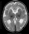
Figure 1
Figure 1
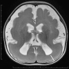
Complete agyria in a DCX mutation patient (Grade 1 on the severity scale). T2-weighted axial MRI image. Wide shallow sylvian fissures create a figure-of-eight appearance. The thickness of the cortex is over 10 mm. A high-intensity (white) line (arrow heads) beneath the cerebral surface is consistent with a cell sparse layer of the four-layered cortex.
Figure 2
Figure 2
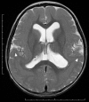
Anterior pachygyria and posterior agyria in a LIS1 mutation patient (Grade 3 on the severity scale). T2-weighted axial MRI image. Note the difference in the width of gyri, the depth of sulci and the thickness of the cortex (bars) between anterior and posterior regions.
Figure 3
Figure 3
Subcortical band heterotopia or double cortex syndrome in a DCX mutation patient (Grade 5 on the severity scale). T2-weighted axial MRI image. Subcortical heterotopic gray matter in the posterior region fuses into the pachygyric cortex in the anterior region (arrowheads).
Figure 4
Figure 4
Schematic diagram of cortical layers in the lissencephaly spectrum compared to the normal brain. Deep cellular layer of the pachygyric or agyric cortex fuses with laminar or band heterotopia in the subcortical white matter, but not with normal six-layered cortex.
Figure 5
Figure 5
Complete agyria in a TUBA1A mutation patient (Grade 1 on the severity scale). T2-weighted axial MRI image (left) and midsagittal image (right). The boundary of the caudate nucleus and lentiform nucleus is obscure. Complete agenesis of the corpus callosum and pontocerebellar hypoplasia are also seen.

References

    1. Abdollahi M. R., Morrison E., Sirey T., Molnar Z., Hayward B. E., Carr I. M., et al. . (2009). Mutation of the variant alpha-tubulin TUBA8 results in polymicrogyria with optic nerve hypoplasia. Am. J. Hum. Genet. 85, 737–744. 10.1016/j.ajhg.2009.10.007 - DOI - PMC - PubMed
    1. Alkuraya F. S., Cai X., Emery C., Mochida G. H., Al-Dosari M. S., Felie J. M., et al. . (2011). Human mutations in NDE1 cause extreme microcephaly with lissencephaly [corrected]. Am. J. Hum. Genet. 88, 536–547. 10.1016/j.ajhg.2011.04.003 - DOI - PMC - PubMed
    1. Bahi-Buisson N., Poirier K., Boddaert N., Saillour Y., Castelnau L., Philip N., et al. . (2008). Refinement of cortical dysgeneses spectrum associated with TUBA1A mutations. J. Med. Genet. 45, 647–653. 10.1136/jmg.2008.058073 - DOI - PubMed
    1. Bahi-Buisson N., Poirier K., Fourniol F., Saillour Y., Valence S., Lebrun N., et al. . (2014). The wide spectrum of tubulinopathies: what are the key features for the diagnosis? Brain 137, 1676–1700. 10.1093/brain/awu082 - DOI - PubMed
    1. Bahi-Buisson N., Souville I., Fourniol F. J., Toussaint A., Moores C. A., Houdusse A., et al. . (2013). New insights into genotype-phenotype correlations for the doublecortin-related lissencephaly spectrum. Brain 136, 223–244. 10.1093/brain/aws323 - DOI - PMC - PubMed