

Subscapularis Tendon Repair Using Suture Bridge Technique
- PMID: 26052489
- PMCID: PMC4454814
- DOI: 10.1016/j.eats.2014.11.013
Subscapularis Tendon Repair Using Suture Bridge Technique
Abstract
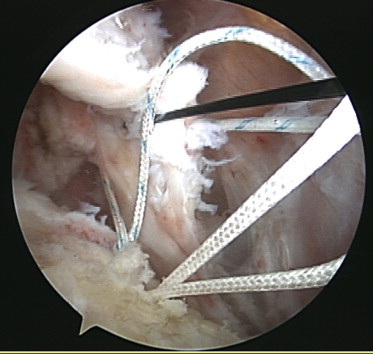
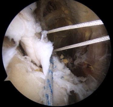
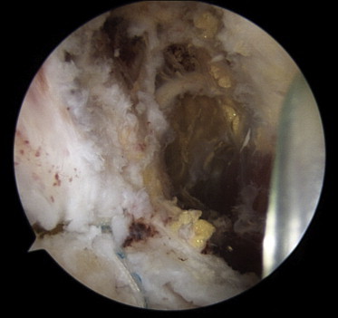
The subscapularis tendon plays an essential role in shoulder function. Although subscapularis tendon tears are less common than other rotator cuff tears, tears of the subscapularis tendon have increasingly been recognized with the advent of magnetic resonance imaging and arthroscopy. A suture bridge technique for the treatment of posterosuperior rotator cuff tears has provided the opportunity to improve the pressurized contact area and mean footprint pressure. However, suture bridge fixation of subscapularis tendon tears appears to be technically challenging. We describe an arthroscopic surgical technique for suture bridge repair of subscapularis tendon tears that obtains ideal cuff integrity and footprint restoration. Surgery using such a suture bridge technique is indicated for large tears, such as tears involving the entire first facet or more, tears with a disrupted lateral sling, and combined medium to large supraspinatus/infraspinatus tears.
Figures


References
-
- Bennett W.F. Arthroscopic repair of isolated subscapularis tears: A prospective cohort with 2- to 4-year follow-up. Arthroscopy. 2003;19:131–143. - PubMed
-
- Adams C.R., Schoolfield J.D., Burkhart S.S. The results of arthroscopic subscapularis tendon repairs. Arthroscopy. 2008;24:1381–1389. - PubMed
-
- Sakurai G., Ozaki J., Tomita Y., Kondo T., Tamai S. Incomplete tears of the subscapularis tendon associated with tears of the supraspinatus tendon: Cadaveric and clinical studies. J Shoulder Elbow Surg. 1998;7:510–515. - PubMed
-
- Lafosse L., Jost B., Reiland Y., Audebert S., Toussaint B., Gobezie R. Structural integrity and clinical outcomes after arthroscopic repair of isolated subscapularis tears. J Bone Joint Surg Am. 2007;89:1184–1193. - PubMed
-
- Burkhart S.S., Tehrany A.M. Arthroscopic subscapularis tendon repair: Technique and preliminary results. Arthroscopy. 2002;18:454–463. - PubMed
LinkOut - more resources
Full Text Sources
Other Literature Sources
