. 2015 Mar 2;4(2):e91-5.
doi: 10.1016/j.eats.2014.11.010. eCollection 2015 Apr.

The "Labral Bridge": A Novel Technique for Arthroscopic Anatomic Knotless Bankart Repair

Affiliations

The "Labral Bridge": A Novel Technique for Arthroscopic Anatomic Knotless Bankart Repair

Roman C Ostermann et al. Arthrosc Tech. .

Abstract

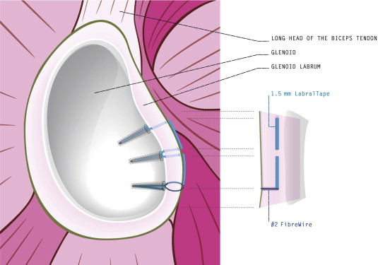
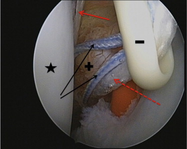
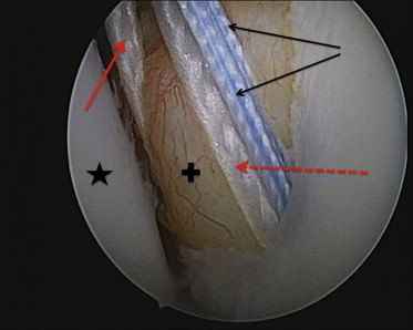
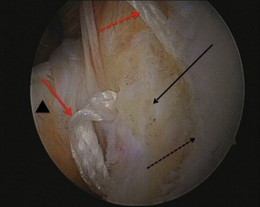
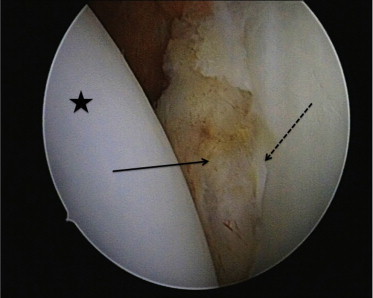
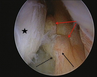
Arthroscopic Bankart repair with suture anchors is widely considered a mainstay for surgical treatment of anterior shoulder instability after recurrent anterior shoulder dislocations. Traditionally, the displaced capsulolabral complex is restored and firmly attached to the glenoid by placing multiple suture anchors individually from a 5- to 3-o'clock position. A variety of different techniques using different anchor designs and materials have been described. Knotless anchors are widely used nowadays for shoulder instability repair, providing a fast and secure way of labral fixation with favorable long-term outcomes. However, these techniques result in a concentrated point load of the reduced labrum to the glenoid at each suture anchor. We describe a technique, developed by the first author, using a 1.5-mm LabralTape (Arthrex, Naples, FL) in combination with knotless suture anchors (3.5-mm PEEK [polyether ether ketone] PushLock anchors; Arthrex), for hybrid fixation of the labrum. The LabralTape is used to secure the torn labrum to the glenoid between each suture anchor, thus potentially providing a more even pressure distribution.


Figures

Fig 1
Fig 1
The LabralTape is secured between the suture anchors, providing an even pressure distribution and potentially encouraging biological healing of the labrum to the glenoid.
Fig 2
Fig 2
Stitch configuration before the first anchor is placed at the 7-o'clock position, viewed from a standard posterior portal in a left shoulder in the beach-chair position. In this case the bone tunnel at the chondrolabral junction is secured with a red plastic stick. The humeral head is marked with a star. Both limbs of the No. 2 FiberWire (black arrows) run through the anchor eyelet (minus sign), in addition to the 1 limb of the LabralTape that runs through the chondrolabral junction (dashed red arrow). The other limb of the LabralTape that leaves the tissue medial to the newly created capsulolabral complex/bump (plus sign) is marked with a solid red arrow.
Fig 3
Fig 3
The first stitch after the anchor has been placed at the 7-o'clock position, viewed from a standard posterior portal in a left shoulder in the beach-chair position. The humeral head is marked with a star, and the newly created capsulolabral complex/bump is marked with a plus sign. Both limbs of the No. 2 FiberWire (black arrows) leave the bone tunnel at the chondrolabral junction together with 1 limb of the LabralTape (dashed red arrow). The other limb of the LabralTape that leaves the tissue medial to the newly created capsulolabral complex/bump (plus sign) is marked with a solid red arrow.
Fig 4
Fig 4
The LabralTape mattress stitch (solid red arrow) running from the second anchor to the desired position of the third anchor before tightening, viewed from a standard posterior portal in a left shoulder in the beach-chair position. The solid black arrow shows the newly created capsulolabral complex/bump, the dashed black arrow shows the chondrolabral junction, and the triangle indicates the subscapularis tendon. The dashed red arrow shows the remaining limb of the LabralTape running out of the anterolateral portal.
Fig 5
Fig 5
The newly created capsulolabral complex/bump (solid arrow) after finalizing the construct, viewed from a standard posterior portal in a left shoulder in the beach-chair position. The dashed arrow marks the chondrolabral junction, and the star indicates the humeral head.
Fig 6
Fig 6
The newly created capsulolabral complex/bump (solid black arrow) after finalizing the construct, viewed from the anterolateral portal. The red arrows mark the grooves within the capsular tissue, created by the LabralTape mattress stitches between the anchors. The dashed black arrow indicates the chondrolabral junction, and the star marks the humeral head. The newly created capsulolabral complex/bump seems well perfused because small vessels can still be observed.

References

    1. Bankart A.S., Cantab M.C. Recurrent or habitual dislocation of the shoulder-joint. 1923. Clin Orthop Relat Res. 1993;(291):3–6. - PubMed
    1. Rowe C.R., Patel D., Southmayd W.W. The Bankart procedure: A long-term end-result study. J Bone Joint Surg Am. 1978;60:1–16. - PubMed
    1. Pope E.J., Ward J.P., Rokito A.S. Anterior shoulder instability—A history of arthroscopic treatment. Bull NYU Hosp Jt Dis. 2011;69:44–49. - PubMed
    1. Cole B.J., Romeo A.A. Arthroscopic shoulder stabilization with suture anchors: Technique, technology, and pitfalls. Clin Orthop Relat Res. 2001;(390):17–30. - PubMed
    1. Ng D.Z., Kumar V.P. Arthroscopic Bankart repair using knot-tying versus knotless suture anchors: Is there a difference? Arthroscopy. 2014;30:422–427. - PubMed

LinkOut - more resources