Review
. 2016 Feb;228(2):233-54.
doi: 10.1111/joa.12314. Epub 2015 Jun 5.

Where hearing starts: the development of the mammalian cochlea

Affiliations
Review

Where hearing starts: the development of the mammalian cochlea

Martin L Basch et al. J Anat. 2016 Feb.

Abstract

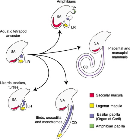
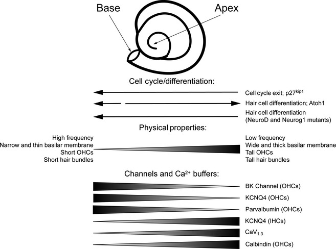
The mammalian cochlea is a remarkable sensory organ, capable of perceiving sound over a range of 10(12) in pressure, and discriminating both infrasonic and ultrasonic frequencies in different species. The sensory hair cells of the mammalian cochlea are exquisitely sensitive, responding to atomic-level deflections at speeds on the order of tens of microseconds. The number and placement of hair cells are precisely determined during inner ear development, and a large number of developmental processes sculpt the shape, size and morphology of these cells along the length of the cochlear duct to make them optimally responsive to different sound frequencies. In this review, we briefly discuss the evolutionary origins of the mammalian cochlea, and then describe the successive developmental processes that lead to its induction, cell cycle exit, cellular patterning and the establishment of topologically distinct frequency responses along its length.

Keywords: BMP; Cochlea; FGF; Hair cells; Notch; Organ of Corti; Sensory; Shh; tonotopy.


Figures

Figure 1
Figure 1
Evolutionary divergence of the inner ear showing the emergence of the cochlea. The aquatic ancestor of modern tetrapods likely had an evagination of the saccule (SA), termed the lagenar recess (LR) that contained the macula lagena (yellow) and a small basilar papilla (purple). This arrangement is seen today in the coelacanth Latimeria (Fritzsch, 1987, 2003) and persists to varying degrees in modern lizards, snakes and turtles, and in many modern amphibians that also have a second unique auditory organ, the amphibian papilla (green). In birds, crocodilians and monotremes, the basilar papilla has elongated to different extents, with the lagenar macula being displaced to the distal tip of the cochlear duct (CD). In therian mammals, the lagena has been lost and the elongated basilar papilla (purple) running the length of the cochlear duct is termed the organ of Corti. In each case, only the pars inferior of the inner ear (saccule, lagenar recess and cochlea duct) are shown in the diagram. This diagram is intended to show the basic trends occurring during the evolution of the cochlea, although in reality considerable variation occurs in the shape and size of the sensory organs in each of the main groups shown in the diagram (Gleich et al. 2004; Manley, 2004, 2012; Smotherman & Narins, 2004; Vater et al. 2004; Fritzsch et al. 2013).
Figure 2
Figure 2
Cardinal axis determination of the amniote inner ear. As the otic placode invaginates to form the otic cup and eventually closes to form the otocyst, the earliest axes established are the M–L axis and the A–P axis, with the D–V axis determined shortly afterwards. A posterior source of RA provides a gradient that allows for the expression of posterior and anterior otic genes. The neural tube provides a source of Wnt to create a dorsalizing gradient, and these dorsalizing signals are augmented by a gradient of inhibitory Gli3R, while the notochord sets up a gradient of Shh, establishing a ventral character. During the otic placode and otic cup stages, Wnt and Fgf3 gradients from the neural tube help establish a medial and a lateral identity.
Figure 3
Figure 3
Development and specification of the prosensory domain (PD) and the organ of Corti (OC). (A) Time course of inner ear development, viewed from the lateral side of the embryo. Between E11.5 and E17.5, the cochlear duct elongates from the ventral wall of the otocyst. Between E12.5 and E13.5, the plates in the vestibular portion undergo extensive rearrangement to form the semicircular canals with cristae (green circles) developing at their base. By E15.5, the vestibular labyrinth and its associated sensory organs have developed (cristae and maculae are shown in green, the approximate position of maculae at E11.5 and E12.5 are shown with red outlines), while the OC continues to differentiate. (B) The bar represents the orientation of the cochlear sections shown in (C). The PD, which gives rise to the OC, is flanked by non‐sensory tissue. On the neural side the PD is flanked by the greater epithelial ridge (GER; red) and on the abneural side by the cells that will form the outer sulcus (blue). (C) Schematic diagrams of cochlear sections at the level of the dotted lines shown in (A). Initially the sensory competent region of the cochlea expresses Sox2 and Jag1. A gradient of BMP4 (visualized by the readout of phosphorylated SMAD1/5/8) from the abneural side of the cochlea refines the prosensory region as development proceeds. By E13.5, cells in the PD exit the cell cycle and contain all the progenitors for the OC. At E14.5, at the border between the GER and the PD, inner hair cells (IHCs) in the base of the cochlea begin to differentiate in response to an as‐yet unidentified signal. As IHCs differentiate, they become a source of FGF8, which together with Notch signaling will help establish the patterning of the OC. By E17.5, the base of the cochlea exhibits its final pattern of one row of IHCs and three rows of outer hair cells (OHCs). Over the next 2 days, this patterning will extend throughout the length of the cochlea. The endolymphatic duct, which grows out from the medial side of the otocyst, is colored purple to distinguish it from the rest of the inner ear. AC, anterior crista ampullaris; ASC, anterior semicircular canal; IPC, inner pillar cell; LC, lateral crista ampullaris; LSC, lateral semicircular canal; OPC, outer pillar cell; PC, posterior crista ampullaris; PSC, posterior semicircular canal; SM, saccular macula; UM, utricular macula.
Figure 4
Figure 4
The structure of the organ of Corti. The organ of Corti rests on the basilar membrane of the cochlear duct. It consists of epithelial cells that are varied in both structure and function. Cochlear hair cells (red) can be anatomically and functionally divided into inner and outer hair cells. The inner hair cells detect sound and transmit the information to the brain via the afferent nerves of the cochleovestibular (VIIIth) ganglion, while outer hair cells are important in amplification of sound and receive efferent innervation from the brainstem. Each hair cell is surrounded by dedicated kinds of supporting cells: inner phalangeal and border cells (light green and gray, respectively) surround the inner hair cells, and Deiters’ cell (green) surround the outer hair cells. In addition, an inner and outer pillar cell (yellow) separate these two domains and form the tunnel of Corti (white area between the pillar cells). Immediately next to the Deiters’ cell are the Hensen's cells (sky‐blue), which have been proposed to modulate the interaction between outer hair cells and the tectorial membrane. The tectorial membrane is an acellular sheet secreted largely by the inner sulcus cells (light purple) in the greater epithelial ridge. Next to the Hensen's cells are the Claudius’ cells (light blue). Cells lying beneath Claudius’ cells and the basilar membrane are known as Boettcher's cells (dark blue). These two types of cells have been reported to maintain the microenvironment of the cochlea, such as Na+ absorption or nitric oxide secretion.
Figure 5
Figure 5
Basal‐to‐apical differences in patterns of cell cycle exit, differentiation and tonotopic properties of the cochlea. Prosensory progenitors exit the cell cycle at the base of the cochlea and express p27kip1 protein in an apical‐to‐basal gradient. However, differentiation of hair cell progenitors begins close to the base of the cochlear duct and spreads down to the apex over the next 4–5 days. This uncoupling of cell cycle exit and differentiation is abolished in a variety of mouse mutants. The physical properties of the cochlear duct and hair cells also vary along the tonotopic axis (basal: high‐frequency; apical: low‐frequency), and tonotopic gradients of ion channels and calcium buffers have also been observed, either in all hair cells or in inner or outer hair cells (IHCs, OHCs).

References

    1. Abdelalim EM, Emara MM, Kolatkar PR (2014) The SOX transcription factors as key players in pluripotent stem cells. Stem Cells Dev 23, 2687–2699. - PubMed
    1. Adam J, Myat A, Le Roux I, et al. (1998) Cell fate choices and the expression of Notch, Delta and Serrate homologues in the chick inner ear: parallels with Drosophila sense‐organ development. Development 125, 4645–4654. - PubMed
    1. Affolter M, Basler K (2007) The Decapentaplegic morphogen gradient: from pattern formation to growth regulation. Nat Rev Genet 8, 663–674. - PubMed
    1. Ahmed M, Wong EY, Sun J, et al. (2012a) Eya1‐Six1 interaction is sufficient to induce hair cell fate in the cochlea by activating Atoh1 expression in cooperation with Sox2. Dev Cell 22, 377–390. - PMC - PubMed
    1. Ahmed M, Xu J, Xu PX (2012b) EYA1 and SIX1 drive the neuronal developmental program in cooperation with the SWI/SNF chromatin‐remodeling complex and SOX2 in the mammalian inner ear. Development 139, 1965–1977. - PMC - PubMed

Publication types

LinkOut - more resources