Noninvasive Computed Tomography-based Risk Stratification of Lung Adenocarcinomas in the National Lung Screening Trial
- PMID: 26052977
- PMCID: PMC4595679
- DOI: 10.1164/rccm.201503-0443OC
Noninvasive Computed Tomography-based Risk Stratification of Lung Adenocarcinomas in the National Lung Screening Trial
Abstract
Rationale: Screening for lung cancer using low-dose computed tomography (CT) reduces lung cancer mortality. However, in addition to a high rate of benign nodules, lung cancer screening detects a large number of indolent cancers that generally belong to the adenocarcinoma spectrum. Individualized management of screen-detected adenocarcinomas would be facilitated by noninvasive risk stratification.
Objectives: To validate that Computer-Aided Nodule Assessment and Risk Yield (CANARY), a novel image analysis software, successfully risk stratifies screen-detected lung adenocarcinomas based on clinical disease outcomes.
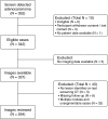
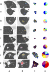
Methods: We identified retrospective 294 eligible patients diagnosed with lung adenocarcinoma spectrum lesions in the low-dose CT arm of the National Lung Screening Trial. The last low-dose CT scan before the diagnosis of lung adenocarcinoma was analyzed using CANARY blinded to clinical data. Based on their parametric CANARY signatures, all the lung adenocarcinoma nodules were risk stratified into three groups. CANARY risk groups were compared using survival analysis for progression-free survival.
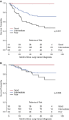
Measurements and main results: A total of 294 patients were included in the analysis. Kaplan-Meier analysis of all the 294 adenocarcinoma nodules stratified into the Good, Intermediate, and Poor CANARY risk groups yielded distinct progression-free survival curves (P < 0.0001). This observation was confirmed in the unadjusted and adjusted (age, sex, race, and smoking status) progression-free survival analysis of all stage I cases.
Conclusions: CANARY allows the noninvasive risk stratification of lung adenocarcinomas into three groups with distinct post-treatment progression-free survival. Our results suggest that CANARY could ultimately facilitate individualized management of incidentally or screen-detected lung adenocarcinomas.
Keywords: image analysis; individualized medicine; lung adenocarcinoma; risk stratification.
Figures


Comment in
-
Noninvasive Quantitative Imaging-based Biomarkers and Lung Cancer Screening.Am J Respir Crit Care Med. 2015 Sep 15;192(6):654-6. doi: 10.1164/rccm.201506-1160ED. Am J Respir Crit Care Med. 2015. PMID: 26371810 Free PMC article. No abstract available.
-
The bell tolls for indeterminant lung nodules: computer-aided nodule assessment and risk yield (CANARY) has the wrong tune.J Thorac Dis. 2016 Aug;8(8):E836-7. doi: 10.21037/jtd.2016.07.85. J Thorac Dis. 2016. PMID: 27618999 Free PMC article. No abstract available.
References
-
- Siegel R, Ma J, Zou Z, Jemal A. Cancer statistics, 2014. CA Cancer J Clin. 2014;64:9–29. - PubMed
-
- Veronesi G, Maisonneuve P, Bellomi M, Rampinelli C, Durli I, Bertolotti R, Spaggiari L. Estimating overdiagnosis in low-dose computed tomography screening for lung cancer: a cohort study. Ann Intern Med. 2012;157:776–784. - PubMed
Publication types
MeSH terms
Grants and funding
LinkOut - more resources
Full Text Sources
Other Literature Sources
Medical

