. 2015 Aug;106(8):1050-6.
doi: 10.1111/cas.12717. Epub 2015 Jul 14.

Class I histone deacetylase inhibitors inhibit the retention of BRCA1 and 53BP1 at the site of DNA damage

Affiliations

Class I histone deacetylase inhibitors inhibit the retention of BRCA1 and 53BP1 at the site of DNA damage

Takayo Fukuda et al. Cancer Sci. 2015 Aug.

Abstract

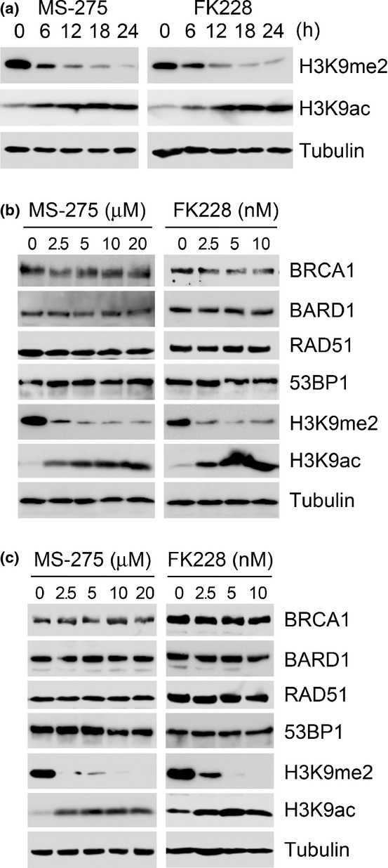
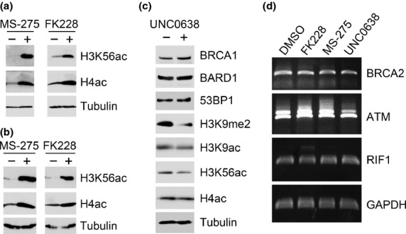
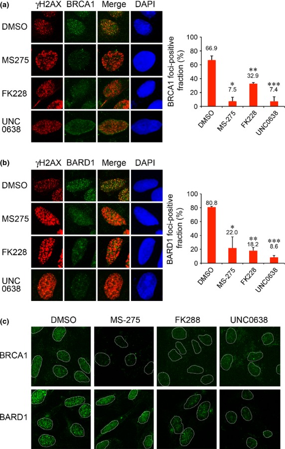
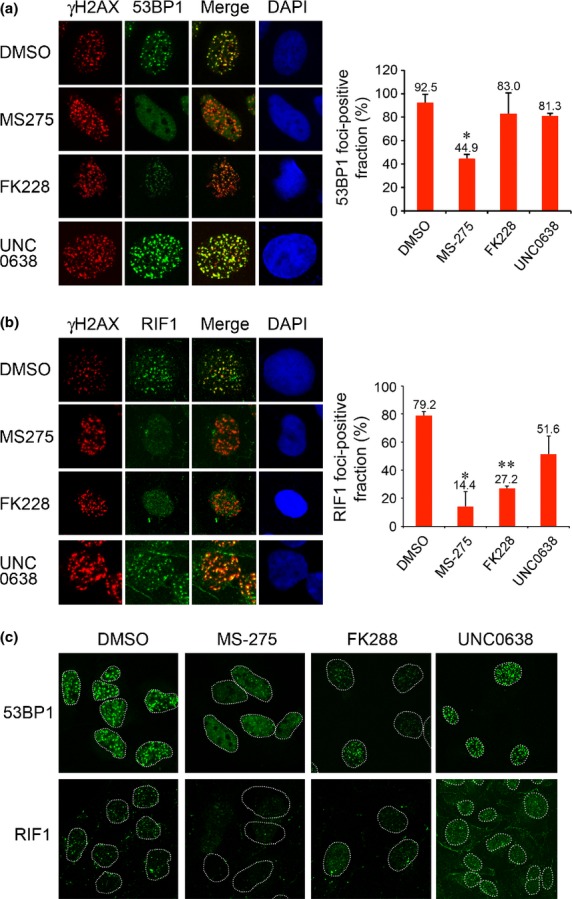
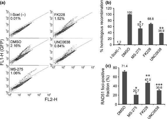
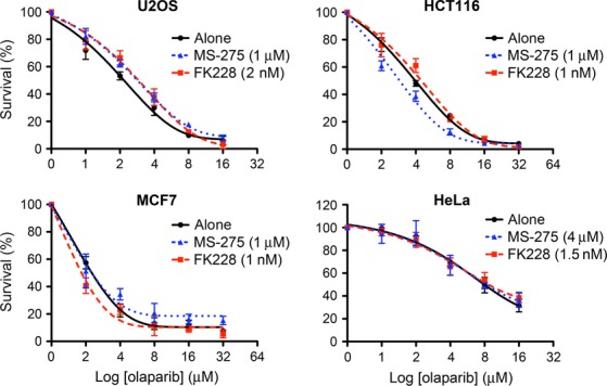
BRCA1 and 53BP1 antagonistically regulate homology-directed repair (HDR) and non-homologous end-joining (NHEJ) of DNA double-strand breaks (DSB). The histone deacetylase (HDAC) inhibitor trichostatin A directly inhibits the retention of 53BP1 at DSB sites by acetylating histone H4 (H4ac), which interferes with 53BP1 binding to dimethylated histone H4 Lys20 (H4K20me2). Conversely, we recently found that the retention of the BRCA1/BARD1 complex is also affected by another methylated histone residue, H3K9me2, which can be suppressed by the histone lysine methyltransferase (HKMT) inhibitor UNC0638. Here, we investigate the effects of the class I HDAC inhibitors MS-275 and FK228 compared to UNC0638 on histone modifications and the DNA damage response. In addition to H4ac, the HDAC inhibitors induce H3K9ac and inhibit H3K9me2 at doses that do not affect the expression levels of DNA repair genes. By contrast, UNC0638 selectively inhibits H3K9me2 without affecting the levels of H3K9ac, H3K56ac or H4ac. Reflecting their effects on histone modifications, the HDAC inhibitors inhibit ionizing radiation-induced foci (IRIF) formation of BRCA1 and BARD1 as well as 53BP1 and RIF1, whereas UNC0638 suppresses IRIF formation of BRCA1 and BARD1 but not 53BP1 and RIF1. Although HDAC inhibitors suppressed HDR, they did not cooperate with the poly(ADP-ribose) polymerase inhibitor olaparib to block cancer cell growth, possibly due to simultaneous suppression of NHEJ pathway components. Collectively, these results suggest the mechanism by that HDAC inhibitors inhibit both the HDR and NHEJ pathways, whereas HKMT inhibitor inhibits only the HDR pathway; this finding may affect the chemosensitizing effects of the inhibitors.

Keywords: 53BP1; BRCA1; DNA damage response; histone deacetylase inhibitor; histone modifications.


Figures

Figure 1
Figure 1
Alteration of H3K9 modifications induced by the histone deacetylase (HDAC) inhibitors MS-275 and FK228. (a) U2OS cells were incubated with MS-275 (2.5 μM) or FK228 (2.5 nM) for the indicated times. Whole cell lysates were immunoblotted with the indicated antibodies. (b, c) U2OS (b) or MCF-7 (c) cells were incubated for 24 h with the indicated doses of MS-275 or FK228 and immunoblotted with the indicated antibodies. MS-275 or FK228 did not inhibit the expression levels of DNA repair genes BRCA1, BARD1, RAD51 and 53BP1 at the doses tested.
Figure 2
Figure 2
Induction of H3K56ac and H4ac by the histone deacetylase (HDAC) inhibitors, but not the HKMT inhibitor at doses that do not affect the expression levels of DNA repair genes. (a, b) U2OS (a) or MCF-7 (b) cells were incubated for 24 h with DMSO (−), MS-275 (2.5 μM) or FK228 (2.5 nM) and immunoblotted with the indicated antibodies. (c) U2OS cells were incubated for 24 h with DMSO (−) or UNC0638 (3 μM) and immunoblotted with the indicated antibodies. UNC0638 did not inhibit the expression levels of DNA repair genes BRCA1, BARD1 and 53BP1 at the doses tested. (d) U2OS cells were treated with MS-275 (2.5 μM), FK228 (2.5 nM) or UNC0638 (3 μM) for 24 h. Gene expression levels for of BRCA2, ATM, RIF1 and the loading control, GAPDH, were analyzed by RT-PCR.
Figure 3
Figure 3
Histone deacetylase (HDAC) inhibitors suppress IRIF formation of BRCA1 and BARD1. (a) U2OS cells treated with DMSO, MS-275, FK228 or UNC0638 were exposed to IR and immunostained with γH2AX and BRCA1. Right panel: Quantification of the cells displaying more than 30 BRCA1 foci. Error bars, SD of two independent experiments, each based on more than 100 cells. Significant differences were calculated compared to DMSO-treated cells: *P = 0.009, **P = 0.015, ***P = 0.011. (b) U2OS cells treated as in (a) were immunostained with γH2AX and BARD1. Error bars, SD of two independent experiments, each based on more than 60 cells. *P = 0.035, **P = 0.003, ***P = 0.001. (c) Low-magnification views. Nuclei are outlined with dashed lines.
Figure 4
Figure 4
Histone deacetylase (HDAC) inhibitors suppress IRIF formation of 53BP1 and RIF1. (a) U2OS cells treated with DMSO, MS-275, FK228 or UNC0638 were exposed to IR and immunostained with γH2AX and 53BP1. Right panel: Quantification of the cells displaying more than 20 53BP1 foci. Error bars, SD of two independent experiments, each based on more than 40 cells. Significant differences were calculated compared to DMSO-treated cells: *P = 0.013. (b) U2OS cells treated as in (a) were immunostained with γH2AX and RIF1. Error bars, SD of two independent experiments, each based on more than 120 cells. *P = 0.024, **P = 0.002. (c) Low-magnification views. Nuclei are outlined with dashed lines.
Figure 5
Figure 5
Histone deacetylase (HDAC) and HKMT inhibitors suppress homology-directed repair (HDR). (a) Fluorescence-activated cell sorting (FACS) data on HDR of I-SceI-induced DSB in HeLa DR-GFP reporter cells treated with DMSO, MS-275, FK228 or UNC0638. (b) Quantification of the GFP-positive fraction. Averages ± SD normalized to cells with DMSO were derived from two independent experiments. Significant differences were calculated compared to DMSO-treated cells: *P = 0.002, **< 0.001. (c) U2OS cells treated with DMSO, MS-275, FK228 or UNC0638 were exposed to infrared (IR) and immunostained with γH2AX and RAD51 6 h after IR. Quantification of the cells displaying more than 20 RAD51 foci. Error bars represent SD from two independent experiments, each based on more than 100 cells. Significant differences were calculated compared to DMSO-treated cells: *P = 0.006, **P = 0.012, ***P = 0.003.
Figure 6
Figure 6
Histone deacetylase (HDAC) inhibitors do not show synthetic lethality with a PARP inhibitor. U2OS, HCT116, MCF7 or HeLa cells were exposed to the indicated doses of olaparib with DMSO (alone), MS-275 or FK228 and analyzed for clonogenic survival. The data are shown with nonlinear regression fit curves of one phase decay (GraphPad Prism). Averages ± SEM, normalized to cells without olaparib, were derived from triplicate experiments.

References

    1. Spiegel S, Milstien S, Grant S. Endogenous modulators and pharmacological inhibitors of histone deacetylases in cancer therapy. Oncogene. 2011;31:537–51. - PMC - PubMed
    1. Groselj B, Sharma NL, Hamdy FC, Kerr M, Kiltie AE. Histone deacetylase inhibitors as radiosensitisers: effects on DNA damage signalling and repair. Br J Cancer. 2013;108:748–54. - PMC - PubMed
    1. Tuzon CT, Spektor T, Kong X, et al. Concerted activities of distinct H4K20 methyltransferases at DNA double-strand breaks regulate 53BP1 nucleation and NHEJ-directed repair. Cell Rep. 2014;8:430–8. - PMC - PubMed
    1. Tang J, Cho NW, Cui G, et al. Acetylation limits 53BP1 association with damaged chromatin to promote homologous recombination. Nat Struct Mol Biol. 2013;20:317–25. - PMC - PubMed
    1. Hsiao K-Y, Mizzen CA. Histone H4 deacetylation facilitates 53BP1 DNA damage signaling and double-strand break repair. J Mol Cell Biol. 2013;5:157–65. - PubMed

Publication types

MeSH terms