. 2016 Aug;229(2):239-51.
doi: 10.1111/joa.12325. Epub 2015 Jun 5.

Comparative brain morphology of Neotropical parrots (Aves, Psittaciformes) inferred from virtual 3D endocasts

Affiliations

Comparative brain morphology of Neotropical parrots (Aves, Psittaciformes) inferred from virtual 3D endocasts

Julieta Carril et al. J Anat. 2016 Aug.

Abstract

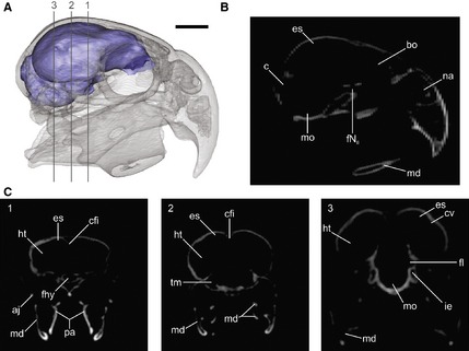
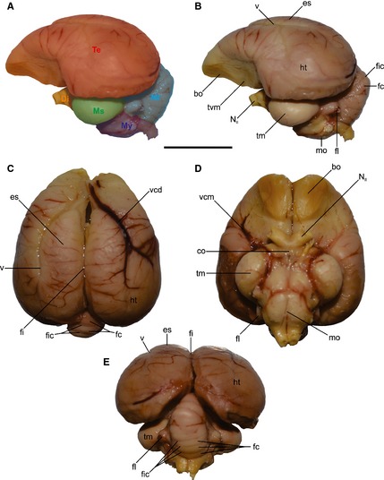
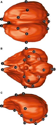
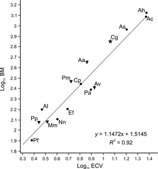
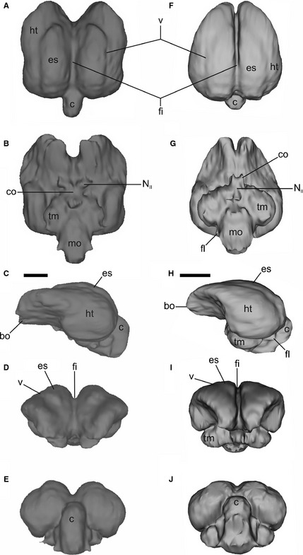
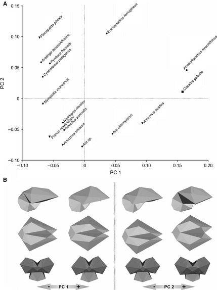
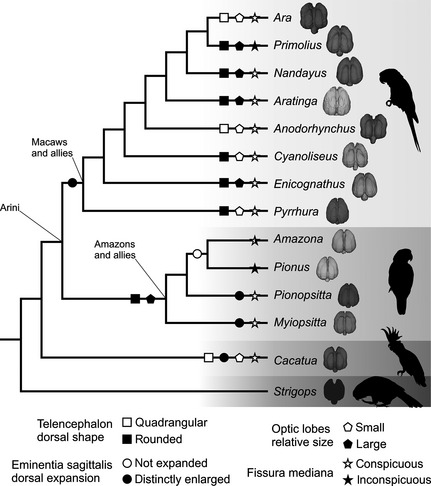
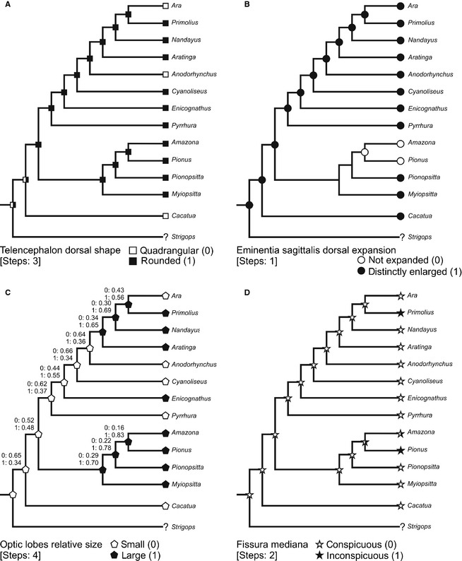
Psittaciformes are a very diverse group of non-passerine birds, with advanced cognitive abilities and highly developed locomotor and feeding behaviours. Using computed tomography and three-dimensional (3D) visualization software, the endocasts of 14 extant Neotropical parrots were reconstructed, with the aim of analysing, comparing and exploring the morphology of the brain within the clade. A 3D geomorphometric analysis was performed, and the encephalization quotient (EQ) was calculated. Brain morphology character states were traced onto a Psittaciformes tree in order to facilitate interpretation of morphological traits in a phylogenetic context. Our results indicate that: (i) there are two conspicuously distinct brain morphologies, one considered walnut type (quadrangular and wider than long) and the other rounded (narrower and rostrally tapered); (ii) Psittaciformes possess a noticeable notch between hemisphaeria that divides the bulbus olfactorius; (iii) the plesiomorphic and most frequently observed characteristics of Neotropical parrots are a rostrally tapered telencephalon in dorsal view, distinctly enlarged dorsal expansion of the eminentia sagittalis and conspicuous fissura mediana; (iv) there is a positive correlation between body mass and brain volume; (v) psittacids are characterized by high EQ values that suggest high brain volumes in relation to their body masses; and (vi) the endocranial morphology of the Psittaciformes as a whole is distinctive relative to other birds. This new knowledge of brain morphology offers much potential for further insight in paleoneurological, phylogenetic and evolutionary studies.

Keywords: 3D brain reconstructions; Aves; character mapping; endocranial morphology; geometric morphometrics.


Figures

Figure 1
Figure 1
Three‐dimensional reconstruction of Cyanoliseus patagonus endocast, generated from CT scans. (A) Lateral view of the skull in transparence showing the endocast; (B, C) tomographs of the skull in (B) sagittal and (C) coronal planes (1–3) showing the spaces occupied by the brain and its different regions/structures. aj, arcus jugalis; bo, bulbus olfactorius; c, cerebellum; cfi, crista frontalis interna; cv, crista vallecularis; es, eminentia sagittalis; fhy, fossa hypohysialis; fl, floculus; fNII, foramen nervus opticum; ht, hemisphaeria telencephali; ie, inner ear; md, mandible; mo, medulla oblongata; na, apertura nasi ossea; pa, ossa palatina; tm, tectum mesencephali. Scale bar: 1 cm.
Figure 2
Figure 2
Dissection of Myiopsitta monachus brain. (A) Regions of the brain in lateral view, (B) left lateral, (C) dorsal, (D) ventral and (E) caudal views showing the main structures of the brain. bo, bulbus olfactorius; co, chiasma opticum; Di, diencephalon; es, eminentia sagittalis; fc, folia cerebeli; fi, fissura interhemispherica; fic, fissure cerebeli; fl, floculus; ht, hemisphaeria telencephalica; mo, medulla oblongata; Ms, mesencephalon; Mt, metencephalon; My, myelencephalon; NII, nervus opticum; Te, telencephalon; tm, tectum mesencephali; tvm, tuber ventromediale; v, vallecula; vcd, vena cerebralis dorsorostralis; vcm, vena cerebralis media. Scale bar: 1 cm.
Figure 3
Figure 3
The 3D brain landmarks used for shape analysis shown in Enicognathus ferrugineus brain. (A) Dorsal, (B) ventral, (C) lateral view. See Table 2 for landmark description.
Figure 4
Figure 4
Scatterplot of log‐transformed brain volume vs. body mass in parrots. Solid star indicates cockatoos; solid dots indicate Macaws and allies; and inverted solid triangles indicate Amazons and allies, according to Tavares et al. (2006). Aa, Amazona aestiva; Av, Amazona vinacea; Ah, Anodorhynchus hyacinthinus; Ac, Ara chloropterus; As, Ara sp.; Al, Aratinga leucophthalma; Cg, Cacatua galerita; Cp, Cyanoliseus patagonus; Ef, Enicognathus ferrugineus; Mm, Myiopsitta monachus; Nn, Nandayus nenday; Pp, Pionopsitta pileata; Pm, Pionus maximiliani; Pa, Primolius auricollis; Pf, Pyrrhura frontalis.
Figure 5
Figure 5
Endocast of adult Anodorhynchus hyacinthinus (A–E) and Pionus maximiliani (F–J) in dorsal, ventral, left lateral, cranial and caudal views. bo, bulbus olfactorius; c, cerebellum; co, chiasma opticum; es, eminentia sagittalis; fi, fissura interhemispherica; fl, floculus; ht, hemispherium telencephali; mo, medulla oblongata; NII, nervus opticum; tm, tectum mesencephali; v, vallecula. Scale bar: 1 cm.
Figure 6
Figure 6
Principle component (PC) analysis of the brain shape of parrots. (A) Plot of PC1 against PC2. (B) Brain shape variation along PC1 and PC2 axis. Solid dots indicate Macaws and allies; inverted solid triangles indicate Amazons and allies; and solid squares indicate cockatoos, according to Tavares et al. (2006).
Figure 7
Figure 7
Phylogenetic diagram plotting the occurrence of some brain morphological traits in Neotropical parrots. Phylogenetic hypothesis modified from Tavares et al. (2006). Characters from Smith & Clarke (2012).
Figure 8
Figure 8
Ancestral‐state reconstruction of endocast in Arini (Psittaciformes) based on Parsimony analysis (steps are indicated between brackets). Additionally in (C), Maximum Likelihood state inferences are indicated by numbers. Characters were taken from Smith & Clarke (2012) and considered as unordered. Phylogenetic proposal modified from Tavares et al. (2006).

References

    1. Balanoff AM, Bever GS, Rowe TB, et al. (2013) Evolutionary origins of the avian brain. Nature 501, 93–97. - PubMed
    1. Baumel JJ, Witmer LM (1993) Osteologia In: Handbook of Avian Anatomy: Nomina Anatomica Avium, No. 23. (eds Baumel J, King A, Breazile J, Evans H, Vanden Berge J.), pp. 45–132. Cambridge, MA: Publications of the Nuttall Ornithological Club.
    1. Bookstein FL (1991) Morphometric Tools for Landmark Data: Geometry and Biology. New York, NY: Cambridge University Pres.
    1. Breazile J, Kuenzel W (1993) Systema nervosum centrale In: Handbook of Avian Anatomy: Nomina Anatomica Avium, No. 23. (eds Baumel J, King A, Breazile J, Evans H, Vanden Berge J.), pp. 493–554. Cambridge, MA: Publications of the Nuttall Ornithological Club.
    1. Carril J, Degrange FJ, Tambussi CP (2015) Jaw myology and bite force of the monk parakeet (Aves, Psittaciformes) Journal of Anatomy: in press. - PMC - PubMed

Publication types

LinkOut - more resources