. 2015 Jul 1;125(7):2690-701.
doi: 10.1172/JCI81589. Epub 2015 Jun 8.

Harnessing endogenous stem/progenitor cells for tendon regeneration

Harnessing endogenous stem/progenitor cells for tendon regeneration

Chang H Lee et al. J Clin Invest. .

Abstract

Current stem cell-based strategies for tissue regeneration involve ex vivo manipulation of these cells to confer features of the desired progenitor population. Recently, the concept that endogenous stem/progenitor cells could be used for regenerating tissues has emerged as a promising approach that potentially overcomes the obstacles related to cell transplantation. Here we applied this strategy for the regeneration of injured tendons in a rat model. First, we identified a rare fraction of tendon cells that was positive for the known tendon stem cell marker CD146 and exhibited clonogenic capacity, as well as multilineage differentiation ability. These tendon-resident CD146+ stem/progenitor cells were selectively enriched by connective tissue growth factor delivery (CTGF delivery) in the early phase of tendon healing, followed by tenogenic differentiation in the later phase. The time-controlled proliferation and differentiation of CD146+ stem/progenitor cells by CTGF delivery successfully led to tendon regeneration with densely aligned collagen fibers, normal level of cellularity, and functional restoration. Using siRNA knockdown to evaluate factors involved in tendon generation, we demonstrated that the FAK/ERK1/2 signaling pathway regulates CTGF-induced proliferation and differentiation of CD146+ stem/progenitor cells. Together, our findings support the use of endogenous stem/progenitor cells as a strategy for tendon regeneration without cell transplantation and suggest this approach warrants exploration in other tissues.


Figures

Figure 7
Figure 7. Tendon healing process by CTGF and endogenous CD146+ TSCs.
Our data collectively suggest that CTGF-treated CD146+ TSCs undergo a robust proliferation phase in 2–7 days of CTGF-initiated healing via the FAK/ERK1/2 signaling pathway. Then, the CD146+ TSCs differentiated into tenocyte-like cells starting at 7 days, which presumably is stimulated by secreted CTGF from CD146 tendon cells. The CD146+ TSCs differentiated by CTGF consequently led to the tendon regeneration featured by reorganized collagen matrix, normal level of cellularity, and functional restoration.
Figure 6
Figure 6. Signaling study in CTGF-treated CD146+ tendon cells.
Western Blot shows that CTGF treatment initiated FAK and ERK1/2 signaling (A). FAK and ERK1/2 were successfully KD using 100 nM Silencer siRNA with Neon system (Invitrogen) (B). FAK and ERK1/2 KD significantly attenuated the proliferation of CD146+ cells promoted by CTGF (C). FAK and ERK1/2 KD attenuated the elevated tendon-related gene expression by CTGF — including Col1a1, Col3a1, Tnc, Vim, and Scx — by 1 week. However, Tnmd expression was elevated with ERK1/2 KD (D). Control (Ctrl) indicates scrambled siRNA (n = 6 biological replicates per each group; *P < 0.01 compared with negative control). One-way ANOVA with post-hoc Tukey HSD was performed. All data are presented as mean ± SD.
Figure 5
Figure 5. Proliferation and differentiation of CD146+ cells in vivo tendon healing.
Ki67+ proliferative CD146+ cells were significantly increased with CTGF delivery by 2 days in comparison with fibrin alone, but there was no difference at 1 week (A). Starting at 1 week, a fraction of CD146+ cells was observed to have be spindle-shaped with aligned collagen fibers in the CTGF-delivered group (B), suggesting that CD146+ cells may undergo tenogenic differentiation. The spindle-shaped tenocyte-like cells derived from CD146+ cells co-expressed COL1A1 and SCX, as demonstrated by immunofluorescence (CE). (n = 10 randomly selected slides per group; *P < 0.001 compared with control without CTGF.) One-way ANOVA with post-hoc Tukey HSD was performed. All data are presented as mean ± SD. White arrows indicate CD146+ perivascular cells; yellow arrowheads indicate spindle-shaped tenocyte-like CD146+ cells.
Figure 4
Figure 4. Tenogenic differentiation of CD146+ tendon cells.
CD146+ tendon cells showed increased collagen deposition upon CTGF treatment, compared with CD146 cells (AC). Expression of tendon/ligament fibroblasts–related genes — including Col1a1, Col3a1, Tnc, Vim, Tnmd, and Scx — was drastically elevated in CD146+ cells upon CTGF treatment in comparison with CD146 cells (D) (n = 6 biological replicates per group; *P < 0.05 compared with control. One-way ANOVA with post-hoc Tukey HSD was performed. All data are presented as mean ± SD. Scale bars: 100 μm.
Figure 3
Figure 3. Fraction of CD146+ tendon cells in vivo upon transection, followed by CTGF delivery.
At 2 days and 1 week, CTGF increased the number of CD146+ cells in the healing region (D and E), compared with without CTGF (A and B). However, the number of CD146+ cells was decreased by 2 weeks both with and without CTGF delivery (C and F). The number of blood vessels increased by 1 week after transection and decreased by 2 weeks (GL). There was no obvious difference in blood vessel number with or without CTGF delivery (GL). Scale bars: 200 μm.
Figure 2
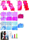
Figure 2. CTGF-enhanced PT healing: H&E in low magnification, high magnification, and Masson’s trichrome.
Low-magnification H&E shows scar-like tissue formation in the healing region (HR) without CTGF (A and C) whereas the CTGF-delivered group promoted healing (B and D) by 2 weeks. Arrows indicate uninjured tendon regions. Consistently in higher magnification, inflammatory matrix with high cell numbers was formed without CTGF (E and L), whereas CTGF attenuated inflammation (H and O) at 2 days. By 1 week, CTGF induced dense alignment of collagen fibers (I and P), in contrast to collagen-lacking scar tissue formed without CTGF (F and M). Native-like highly aligned collagen fibers were formed after 4 weeks CTGF delivery (J and Q), in contrast to scar-like matrix without CTGF (G and N). Native PT sections are shown in K and R. Furthermore, tensile stiffness of healed tendons with CTGF delivery was significantly higher than without CTGF and corresponded to that of native tissue (S and T) (n = 6 tissue samples per group; *P = 0.016 and 0.019 compared with +CTGF and native, respectively). One-way ANOVA with post-hoc Tukey HSD was performed. All data are presented as mean ± SD.
Figure 1
Figure 1. CD146+ cells in tendons.
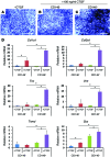
Immunofluorescence revealed CD146+ cells surrounding blood vessels in rat PT (A and B). Flow cytometry showed that approximately 0.8% of isolated cells from rat PT are highly positive for CD146 (C). Approximately 1% of adherent MNCs from PT were CD146+ (D and F), whereas approximately 72% of CFU-F were CD146+ (n = 6 per group, P < 0.0001) (E and F). Treatment with 100 ng/ml of CTGF significantly increased the number of CD146+ cells (GI). In addition, in vitro fate of CD146 expression was regulated by CTGF. CTGF treatment for 4 weeks failed to induce any CD146 expression in the sorted CD146 tendon cells (JL). Sorted CD146+ tendon cells maintained CD146 expression with CTGF treatment by 4 weeks (M and N). However, CD146 expression was diminished in CD146+ tendon cells cultured for 4 weeks without CTGF treatment (O). All data are presented as mean ± SD. Images were selected as the representatives of 6 replicates total.

References

    1. Arany PR, et al. Photoactivation of endogenous latent transforming growth factor-beta1 directs dental stem cell differentiation for regeneration. Sci Transl Med. 2014;6(238):238ra269. - PMC - PubMed
    1. Chen FM, Wu LA, Zhang M, Zhang R, Sun HH. Homing of endogenous stem/progenitor cells for in situ tissue regeneration: Promises, strategies, and translational perspectives. Biomaterials. 2011;32(12):3189–3209. doi: 10.1016/j.biomaterials.2010.12.032. - DOI - PubMed
    1. Miller FD, Kaplan DR. Mobilizing endogenous stem cells for repair and regeneration: are we there yet? Cell Stem Cell. 2012;10(6):650–652. doi: 10.1016/j.stem.2012.05.004. - DOI - PubMed
    1. Vanden Berg-Foels WS. In situ tissue regeneration: chemoattractants for endogenous stem cell recruitment. Tissue Eng Part B Rev. 2014;20(1):28–39. doi: 10.1089/ten.teb.2013.0100. - DOI - PMC - PubMed
    1. Fodor WL. Tissue engineering and cell based therapies, from the bench to the clinic: the potential to replace, repair and regenerate. Reprod Biol Endocrinol. 2003;1:102. doi: 10.1186/1477-7827-1-102. - DOI - PMC - PubMed

MeSH terms