Review
. 2015 Jun 8;7(8):a020529.
doi: 10.1101/cshperspect.a020529.

Schwann cell myelination

Affiliations
Review

Schwann cell myelination

James L Salzer. Cold Spring Harb Perspect Biol. .

Abstract

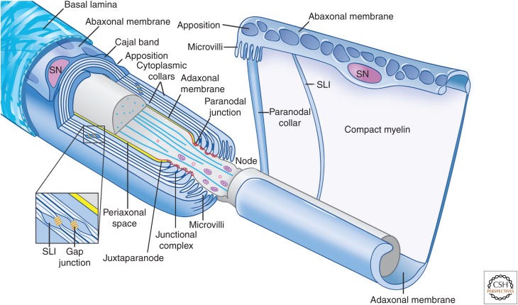
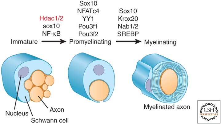
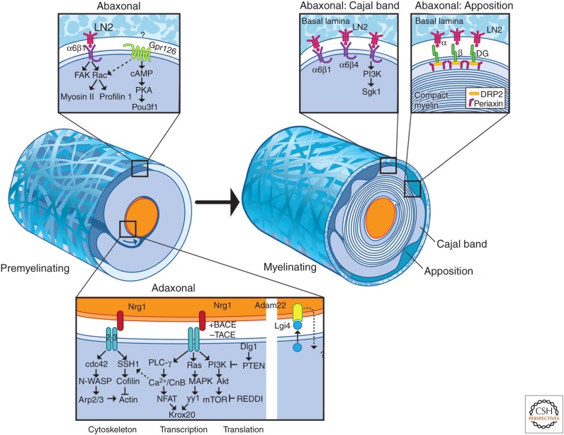
Myelinated nerve fibers are essential for the rapid propagation of action potentials by saltatory conduction. They form as the result of reciprocal interactions between axons and Schwann cells. Extrinsic signals from the axon, and the extracellular matrix, drive Schwann cells to adopt a myelinating fate, whereas myelination reorganizes the axon for its role in conduction and is essential for its integrity. Here, we review our current understanding of the development, molecular organization, and function of myelinating Schwann cells. Recent findings into the extrinsic signals that drive Schwann cell myelination, their cognate receptors, and the downstream intracellular signaling pathways they activate will be described. Together, these studies provide important new insights into how these pathways converge to activate the transcriptional cascade of myelination and remodel the actin cytoskeleton that is critical for morphogenesis of the myelin sheath.


Figures

Figure 1.
Figure 1.
Organization of myelinating Schwann cells. Schematic organization of myelinating Schwann cells (blue) surrounding an axon (gray); the left cell is shown in longitudinal cross section and the right cell is shown unwrapped. Myelinating Schwann cells are surrounded by a basal lamina (illustrated only on the left), which is in direct contact with the abaxonal membrane. The abaxonal compartment contains the Schwann cell nucleus (SN); it is divided into Cajal bands and periodic appositions that form between the abaxonal membrane and outer turn of compact myelin. The Schwann cell adaxonal membrane is separated from the axonal membrane by the periaxonal space (shown in yellow). Compact myelin is interrupted by Schmidt–Lanterman incisures (SLI), which retain cytoplasm and are enriched in the gap and other junctions; a similar autotypic junctional complex of adherens, tight and gap junctions, forms between the apposed membranes of the paranodal loops. Also shown are the paranodal loops and junctions (red) and the Schwann cell microvilli contacting the axon at the node. The axon diameter is reduced in the region of the node and paranodes. (This figure is adapted, with permission, from an original figure in Salzer 2003; modified in Nave 2010.)
Figure 2.
Figure 2.
Cajal bands. Fluorescence micrograph showing a portion of a teased, myelinated fiber from adult rat sciatic nerve, the presence of the Cajal bands (green), and the membrane appositions (red) in the abaxonal compartment. Staining shown is for phospho-NDRG1 (green) and α-dystroglycan (red) (see Heller et al. 2014 for further details).
Figure 3.
Figure 3.
Schematic organization of the compact myelin sheath. Myelin forms, initially, as loose wraps of uncompacted membrane. With the onset of myelin transcription, myelin proteins are up-regulated, including P0, maltose-binding protein (MBP), and PMP22, which are shown diagrammatically. Compaction is mediated by extracellular interactions of P0 tetramers on one membrane interacting with P0 tetramers on the opposing membrane, shown here only as dimer/dimer interactions for simplicity. Compaction of the cytoplasmic leaflets is mediated by electrostatic interactions of MBP with the phospholipid bilayer supplemented by the interactions with the cytoplasmic tail of P0. Compact myelin appears on electron microscopy (EM) as a major dense line (MDL) (representing tight apposition of the cytoplasmic leaflets) alternating with the intraperiod lines (representing the two apposed extracellular leaflets) as shown.
Figure 4.
Figure 4.
Transcriptional cascade of myelination. Expression of TFs at different stages of the Schwann cell lineage is shown. At the immature stage, expression of Sox10 and of Hdac1/2 (epigenetic regulators, red) is essential for progression to the promyelinating stage; NF-κB promotes, but is not essential, during the Schwann cell lineage. Progression from the promyelinating to the myelinating phenotype requires Pou3f1 (Oct6) and Pou3f2 (Brn2) for timely advance; Sox10, NFATc4, and YY1 are essential. These latter proteins all promote expression of Krox20, which, together with the interacting Nab proteins, is essential for the expression of the myelinating phenotype. Lipid biosynthesis is up-regulated via SREBP.
Figure 5.
Figure 5.
Schematic of extrinsic signals, receptors, and intracellular signaling pathways that regulate myelination. This figure summarizes the key extrinsic signals, their receptors, and the downstream signaling pathways active in the adaxonal and abaxonal compartments. The major axonal signals include type III NRG1 and Adam22 (disintegrin and metalloprotease), which signal via erbB receptors and Lgi4 (leucine-rich glioma inactivated), respectively. NRG1 is subject to protease cleavage that is activating (BACE, β-secretase) or inactivating (TACE, tumor necrosis factor–α-converting enzyme). Major pathways downstream from erbB signaling include (1) phospholipase C (PLC)-γ, calcineurin B (CnB), and nuclear factor of activated T cells (NFAT), (2) mitogen-activated protein kinase (MAPK), and (3) PI3K, Akt, and the mammalian target of rapamycin (mTOR). NFATc4 and YY1 drive transcription of Krox20; mTOR is a regulator of cap-dependent protein synthesis. NRG signaling also drives the remodeling of the actin cytoskeleton as shown. In the abaxonal compartment before myelination, laminin signaling activates FAK and Rac to promote radial sorting. Gpr126 regulates cAMP and protein kinase A (PKA) to promote sorting and myelination; its assignment to the abaxonal compartment is tentative and its ligand(s) at the time of this review has not been reported. With maturation, the abaxonal compartment organizes into the cytoplasmic channels, termed Cajal bands, and membrane appositions. Signaling in the Cajal bands is mediated in part via integrins. The membrane apposition is mediated by a complex of dystroglycan, DRP2, and periaxin; the space between the baseline (BL) and the appositions as shown is exaggerated for artistic purposes. See the text for additional details on these pathways. N-WASP, Neuronal Wiskott–Aldrich syndrome protein.

References

    1. Abe I, Ochiai N, Ichimura H, Tsujino A, Sun J, Hara Y. 2004. Internodes can nearly double in length with gradual elongation of the adult rat sciatic nerve. J Orthop Res 22: 571–577. - PubMed
    1. Aguayo AJ, Bray GM, Perkins SC. 1979. Axon-Schwann cell relationships in neuropathies of mutant mice. Ann NY Acad Sci 317: 512–531. - PubMed
    1. Armstrong R, Toews AD, Morell P. 1987. Axonal transport through nodes of Ranvier. Brain Res 412: 196–199. - PubMed
    1. Arthur-Farraj P, Wanek K, Hantke J, Davis CM, Jayakar A, Parkinson DB, Mirsky R, Jessen KR. 2011. Mouse Schwann cells need both NRG1 and cyclic AMP to myelinate. Glia 59: 720–733. - PMC - PubMed
    1. Atanasoski S, Scherer SS, Sirkowski E, Leone D, Garratt AN, Birchmeier C, Suter U. 2006. ErbB2 signaling in Schwann cells is mostly dispensable for maintenance of myelinated peripheral nerves and proliferation of adult Schwann cells after injury. J Neurosci 26: 2124–2131. - PMC - PubMed

Publication types