Review
. 2015 Jul;93(7):707-17.
doi: 10.1007/s00109-015-1297-8. Epub 2015 Jun 10.

Autophagy in autoimmune disease

Affiliations
Review

Autophagy in autoimmune disease

Zhen Yang et al. J Mol Med (Berl). 2015 Jul.

Abstract

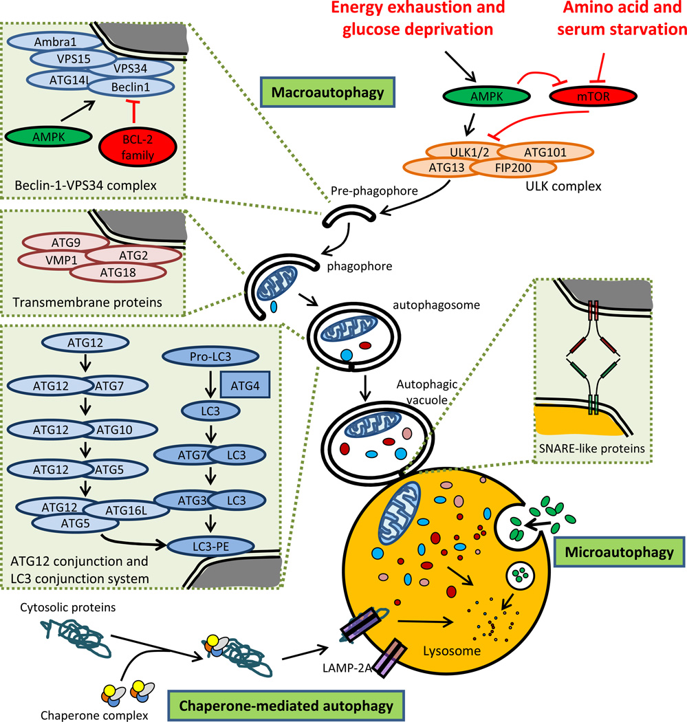
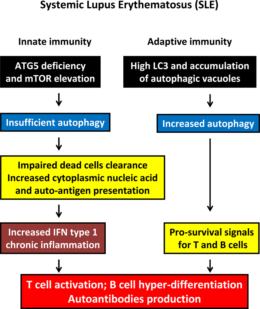
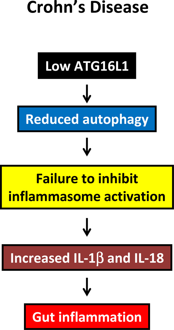
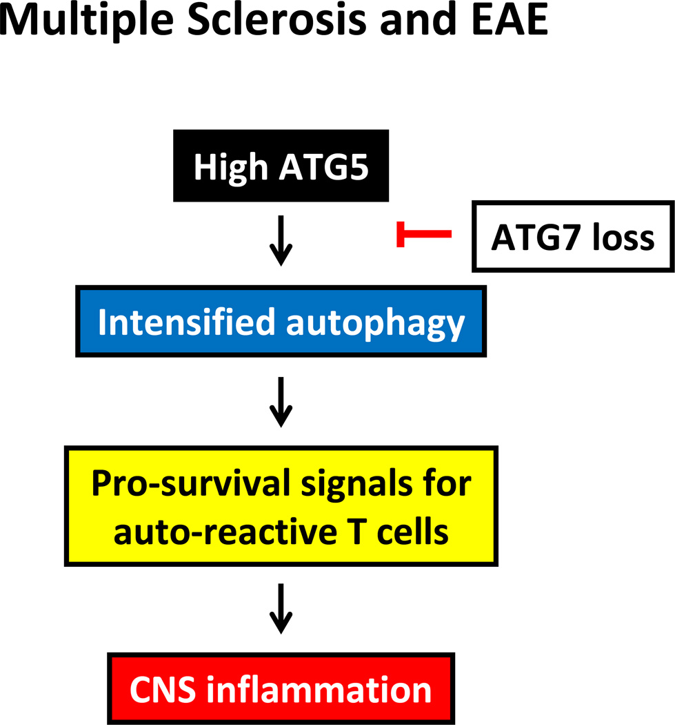
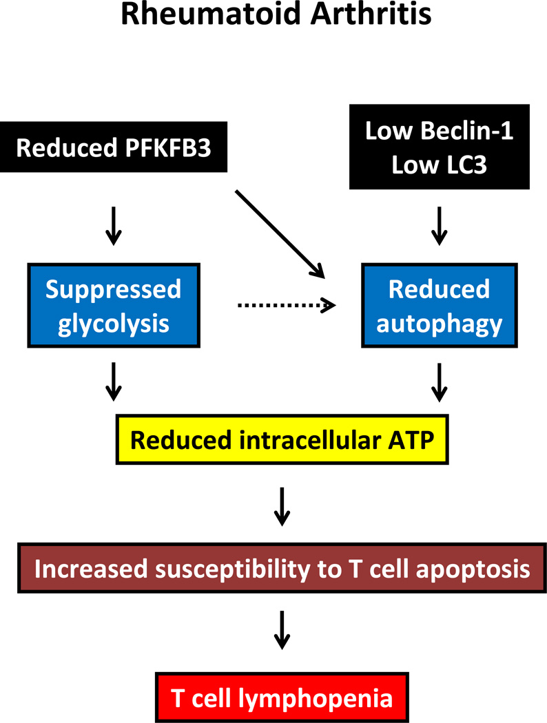
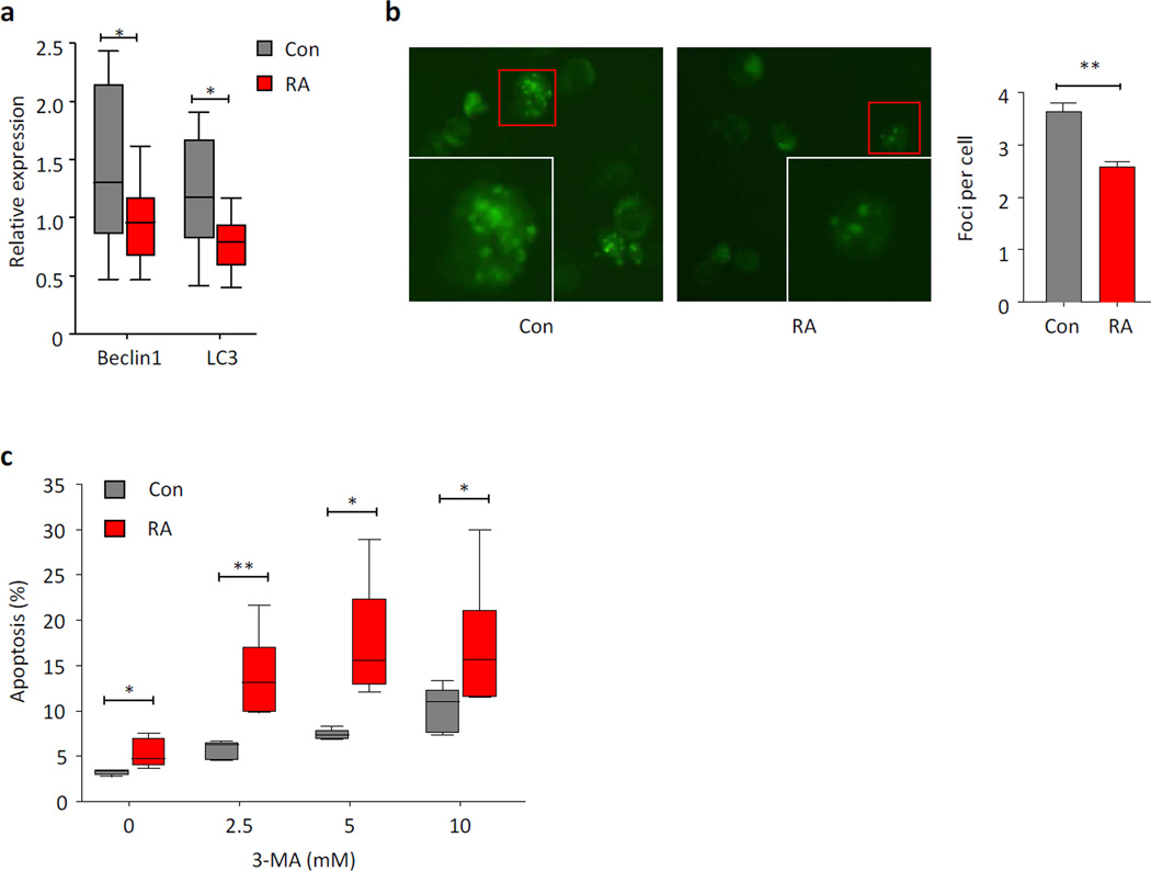
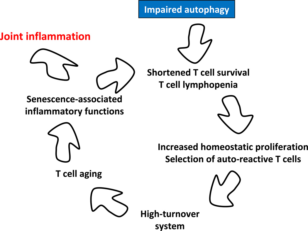
Autophagy is a protective and life-sustaining process in which cytoplasmic components are packaged into double-membrane vesicles and targeted to lysosomes for degradation. This process of cellular self-digestion is an essential stress response and is cytoprotective by removing damaged organelles and proteins that threaten the cell's survival. Key outcomes include energy generation and recycling of metabolic precursors. In the immune system, autophagy regulates processes such as antigen uptake and presentation, removal of pathogens, survival of short- and long-lived immune cells, and cytokine-dependent inflammation. In all cases, a window of optimal autophagic activity appears critical to balance catabolic, reparative, and inflammation-inducing processes. Dysregulation of autophagosome formation and autophagic flux can have deleterious consequences, ranging from a failure to "clean house" to the induction of autophagy-induced cell death. Abnormalities in the autophagic pathway have been implicated in numerous autoimmune diseases. Genome-wide association studies have linked polymorphisms in autophagy-related genes with predisposition for tissue-destructive inflammatory disease, specifically in inflammatory bowel disease and systemic lupus erythematosus. Although the precise mechanisms by which dysfunctional autophagy renders the host susceptible to continuous inflammation remain unclear, autophagy's role in regulating the long-term survival of adaptive immune cells has recently surfaced as a defect in multiple sclerosis and rheumatoid arthritis. Efforts are underway to identify autophagy-inducing and autophagy-suppressing pharmacologic interventions that can be added to immunosuppressive therapy to improve outcomes of patients with autoimmune disease.


Conflict of interest statement

Conflict of interest The authors declare no competing financial interests.

Figures

Figure 1
Figure 1. Schematic diagram of the main autophagic pathways
Autophagy is modulated by environmental nutrient signals via a signaling pathway dependent on AMP-activated protein kinase (AMPK) and the mammalian (mechanistic) target of rapamycin (mTOR). In the absence of amino acids or in response to other stimuli, mTOR negatively regulates UNC-51–like kinase 1 (ULK1) through direct phosphorylation and destabilization of ULK1 protein, thus acting as a negative regulator of autophagy. Under energy depletion, AMPK negatively regulates mTOR and also directly phosphorylates ULK1, thereby acting as a positive regulator of autophagy. Macroautophagy is regulated by a set of autophagy-related proteins (ATG proteins). During macroautophagy, cytoplasmic constituents are engulfed in an isolation membrane that is elongated mainly through the action of two ubiquitin-like conjugation systems into a double-membraned autophagosome. Autophagosomes fuse with lysosomes to form autolysosomes, resulting in complete degradation of the sequestered cytoplasmic components by lysosomal hydrolases. In the case of microautophagy, cytosolic proteins are directly internalized in single membrane vesicles into lysosome by invaginating, protrusion and/or septation. In CMA, specific cytosolic proteins are transported into lysosomes via a molecular chaperone/receptor complex composed of HSPA8/HSC70 and LAMP-2A (the CMA receptor lysosome-associated membrane protein type-2A).
Figure 2
Figure 2. Autophagy in SLE
In SLE, maladaptive autophagy is present in both innate and adaptive immunity. Insufficient autophagy due to the potential involvement of autophagic genes (e.g., Atg5) results in impaired dead cell clearance, increased autoantigen presentation and excessive type I IFN production. Conversely, increased autophagic activity when present in lymphocytes promotes T and B cell survival. Thus, defective clearance of apoptotic cells, increased antigenic load in APCs, excess T cell help, B cell hyperdifferentiation, and increased autoantibody production are all important contributors to the development of SLE.
Figure 3
Figure 3. Autophagy in Crohn’s disease
In Crohn’s disease, SNPs in the autophagic gene ATG16L1 are suspected to cause an autophagy defect that leads to failed inhibition of inflammasome activation and, thereafter, increasing cytokine production and chronic gut inflammation.
Figure 4
Figure 4. Autophagy in CNS inflammation
In multiple sclerosis/EAE, intensified autophagy supports the survival of autoreactive T cells and results in CNS inflammation.
Figure 5
Figure 5. Autophagy in RA
In RA, low expression of autophagic genes, as well as insufficiency of the glycolytic enzyme PFKFB3, leave T cells with low glycolytic flux and ATP deficiency. This metabolic stress confers susceptibility for apoptotic death and eventually results in a state of lymphopenia.
Figure 6
Figure 6. Repressed autophagic activity in RA T cells
CD4 T cells from RA patients fail to upregulate autophagic genes (a) and generate low numbers of GFP-LC3 foci (b) when stimulated through the T cell receptor. Defective autophagosome formation and autophagic flux renders the T cells highly sensitive to the autophagy inhibitor 3-MA (c). The frequency of apoptotic cells is shown in relationship to increasing doses of 3-MA. Cells from RA patients and age-matched controls are compared.
Figure 7
Figure 7. Defective autophagy as a risk factor for autoimmunity – a hypothetical model
In response to antigen and danger signals, lymphocytes mobilize autophagy as a basal mechanism to alleviate stress and fulfill the energy demands associated with clonal expansion. T cells from RA patients fail to upregulate autophagic activity, rendering them susceptible to apoptosis. Evolving lymphopenia triggers compensatory homeostatic proliferation to make up for the T cell loss. Proliferating T cells are selected on autoantigens, biasing the T cell repertoire towards recognition of self. Chronic T cell loss sustains a high-turnover system, eventually causing T cell aging. Prematurely aged T cells acquire the so-called senescence-associated secretory phenotype (SASP) and release massive amounts of inflammatory mediators. The end result is a host predisposed to tissue inflammation. Synovia-specific antigens may direct hyperinflammatory T cells towards the joint.

References

    1. Ravikumar B, Sarkar S, Davies JE, Futter M, Garcia-Arencibia M, Green-Thompson ZW, Jimenez-Sanchez M, Korolchuk VI, Lichtenberg M, Luo S, et al. Regulation of mammalian autophagy in physiology and pathophysiology. Physiol Rev. 2010;90:1383–1435. - PubMed
    1. Klionsky DJ, Emr SD. Autophagy as a regulated pathway of cellular degradation. Science. 2000;290:1717–1721. - PMC - PubMed
    1. Shintani T, Klionsky DJ. Autophagy in health and disease: a double-edged sword. Science. 2004;306:990–995. - PMC - PubMed
    1. Geng J, Klionsky DJ. The Atg8 and Atg12 ubiquitin-like conjugation systems in macroautophagy. 'Protein modifications: beyond the usual suspects' review series. EMBO Rep. 2008;9:859–864. - PMC - PubMed
    1. Fujita N, Itoh T, Omori H, Fukuda M, Noda T, Yoshimori T. The Atg16L complex specifies the site of LC3 lipidation for membrane biogenesis in autophagy. Mol Biol Cell. 2008;19:2092–2100. - PMC - PubMed

Publication types

LinkOut - more resources