. 2015 Jun 9:15:177.
doi: 10.1186/s12906-015-0680-x.

Antifungal activity of berberine hydrochloride and palmatine hydrochloride against Microsporum canis -induced dermatitis in rabbits and underlying mechanism

Affiliations

Antifungal activity of berberine hydrochloride and palmatine hydrochloride against Microsporum canis -induced dermatitis in rabbits and underlying mechanism

Chen-Wen Xiao et al. BMC Complement Altern Med. .

Abstract

Background: Phellodendron amurense, exhibits antifungal activity mainly by bioactive components including berberine hydrochloride and palmatine hydrochloride. This study was conducted to evaluate the antifungal effects of berberine hydrochloride, palmatine hydrochloride, and a mixture of both substances against Microsporum canis in vivo and in vitro.

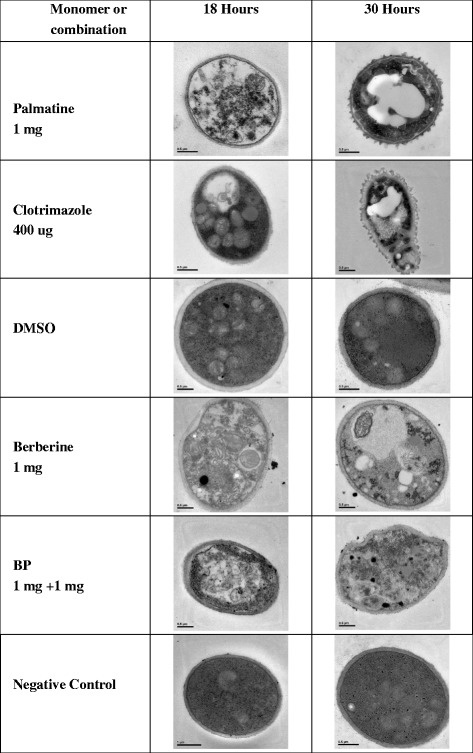
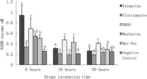
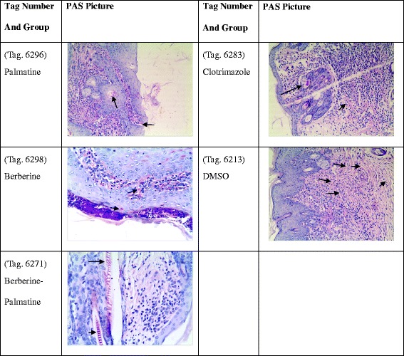
Methods: The minimal inhibitory concentrations (MICs) of monomers and clotrimazole were determined using 1.5 % tryptic soy agar. The effects of these drugs on Microsporum canis growth was detected by determining dry weight. Transmission electron microscopy (TEM) was performed to observe the effect of chemicals on cell ultrastructure. Differential mRNA expressions of eight genes of M. canis treated with berberine or palmatine or their combination at different time points were determined by real-time PCR. NADH enzyme concentration was also detected. Clinical evaluation via in-vivo antifungal assay was also performed. Skin histology PAS staining was also carried out.

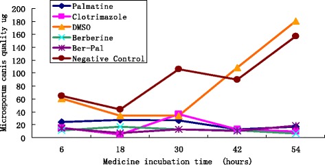
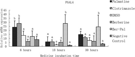
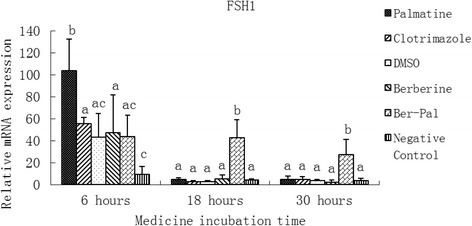
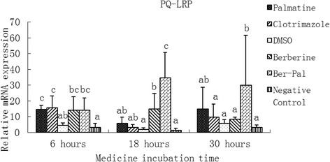
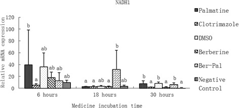
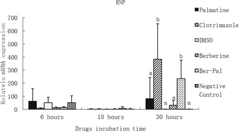
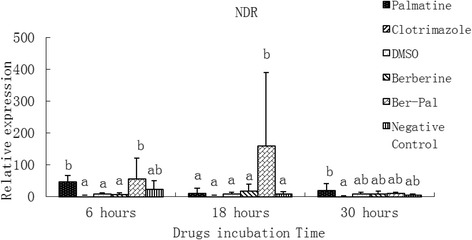
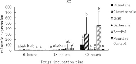
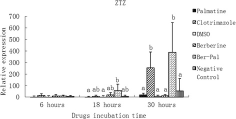
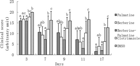
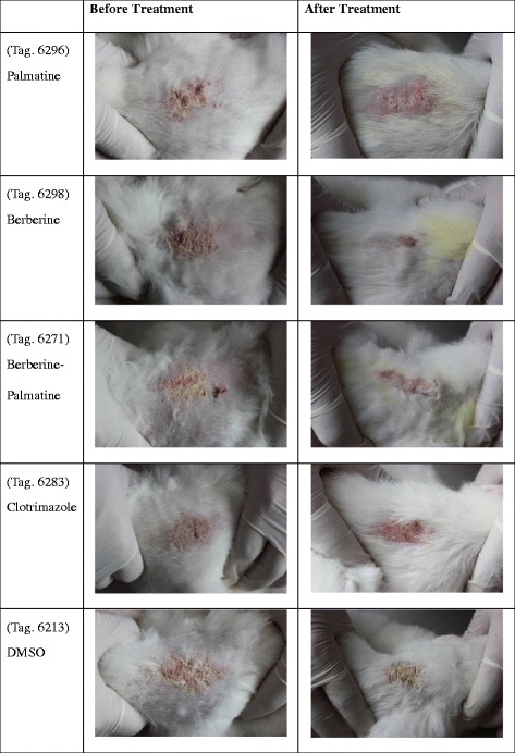
Results: Results showed that MICs of berberine, palmatine and clotrimazole were 1, 1, and 0.015 mg/mL, respectively. No significant difference was observed among the growth curves of the three groups before 18 h was reached. TEM showed that these drugs could destroy the cell membrane and organelles of M. canis at different time points. After 30 h of incubation, relative mRNA expressions of the genes in the combined group were significantly higher than those in the other groups including the clotrimazole group (P < 0.05); Palmatine initially induced the mRNA up-regulation of PGAL4, FSH1, PQ-LRP, NADH1 and NDR in M. canis; by contrast, berberine maintained a high expression level of these genes to shorten fungal life cycle and eradicate M. canis. Clinical results showed that combined treatment was more effective than single administration of each monomer or clotrimazole. Hence, berberine mixed with palmatine significantly elicited antifungal activities and could be used to treat M. canis in rabbits.

Conclusion: These results provide a comprehensive view of the mechanism of berberine and palmatine in anti-M. canis activity.


Figures

Fig. 1
Fig. 1
Effects of berberine hydrochloride and palmatine hydrochloride on the growth of M. canis. M. canis cells were treated for 54 h with berberine hydrochloride and palmatine hydrochloride or their combination at concentrations of 1 mg/mL, 1 mg/mL, 1 mg/mL + 1 mg/mL respectively; clotrimazole at a concentration of 0.4 mg/mL; or DMSO or Negative control
Fig. 2
Fig. 2
Electron microscopic observation on the morphology of M. canis under different medicines or cultivation hours; 1 mg of palmatine or berberine or B-P combination (1 mg + 1 mg) or 400 ug of clotrimazole, DMSO or negative control. The shapes of M. canis in different medicine groups with 18 h were abnormal except DMSO or Negative control group. After 30 h incubation, the degree of damage of M. canis in medicine groups became more serious than 18 h and still there were no changes in DMSO or Negative control group
Fig. 3
Fig. 3
Inhibitory effects of different chemicals on M. canis porphyrin galactose 4 (PGAL4) gene expression. Different letters in the bars show significantly difference (P < 0.05). After 6 h of incubation, the relative mRNA expressions of PGAL4 in the palmatine group and the clotrimazole group were significantly higher than those in other groups (P <0.05). After 18 and 30 h, the relative mRNA expression of PGAL4 in the B-P group was significantly higher than that in other groups (P < 0.05; Fig. 3)
Fig. 4
Fig. 4
Inhibitory effects of different chemicals on M. canis family of serine hydrolases1 (FSH1) gene expression. Different letters in the bars show significantly difference (P <0.05). After 6 h of incubation, relative mRNA expression of FSH1 in the palmatine group was significantly higher than that of the other groups (P < 0.05). After 18 and 30 h, the highest relative mRNA expression of FSH1 was found in the B-P group (P < 0.05)
Fig. 5
Fig. 5
Inhibitory effects of different chemicals on M. canis PQ loop repeat protein (PQ-LRP) gene expression. Different letters in the bars show significantly difference (P <0.05)After 6 h of incubation, the relative mRNA expressions of PQ-LRP in drug-treated groups were significantly higher than those of DMSO and Negative control groups (P < 0.05). After 18 h, the relative mRNA expressions of PQ-LRP in B-P and B groups were significantly higher than those in DMSO and Negative control groups (P < 0.05). The relative mRNA expression of PQ-LRP in the B-P group was also significantly higher than that in the berberine group and the clotrimazole group (P < 0.05). After 30 h, the relative mRNA expression of PQ-LRP in the B-P group was significantly higher than that in the other groups except in the palmatine group (P < 0.05)
Fig. 6
Fig. 6
Inhibitory effects of different chemicals on M. canis NADH dehydrogenase subunit 1 (NADH 1) gene expression. Different letters in the bars show significantly difference (P <0.05)After 6 h of incubation, the relative mRNA expression of NADH1 in the palmatine group was significantly higher than that in the clotrimazole group (P < 0.05). After 18 h, the relative mRNA expression of NADH1 in the B-P group was significantly higher than that in the other groups except DMSO and Negative control groups (P < 0.05). After 30 h, the relative mRNA expressions of NADH1 in palmatine, DMSO and B-P groups were significantly higher than those in the other groups (P < 0.05)
Fig. 7
Fig. 7
Inhibitory effects of different chemicals on M. canis Ribonucleoprotein (RNP) gene expression. Different letters in the bars show significantly difference (P <0.05).After 30 h of incubation, the relative mRNA expressions of RNP in B-P and clotrimazole groups were significantly higher than those in the other groups (P < 0.05)
Fig. 8
Fig. 8
Inhibitory effects of different chemicals on M. canis NADPH-dependent D-xylose reductase (NDR) gene expression. Different letters in the bars show significantly difference (P <0.05).After 6 h of incubation, the relative mRNA expressions of NDR in palmatine and B-P groups were significantly higher than those in the other groups except the Negative control group (P < 0.05). After 18 h, the relative mRNA expression of NDR in the B-P group was significantly higher than that in the other groups (P < 0.05)
Fig. 9
Fig. 9
Inhibitory effects of different chemicals on M. canis symbiotic chitinase (SC) gene expression. Different letters in the bars show significantly difference (P <0.05). After 30 h of incubation, the relative mRNA expressions of SC in B-P and clotrimazole groups were significantly higher than those in the other groups (P < 0.05)
Fig. 10
Fig. 10
Inhibitory effects of different chemicals on M. canis Zinc transporter zupT (ZTZ) gene expression. Different letters in the bars show significantly difference (P <0.05).After 30 h of incubation, the relative mRNA expressions of ZTZ in B-P and clotrimazole groups were significantly higher than those in the other groups (P < 0.05)
Fig. 11
Fig. 11
Levels of M. canis NADH enzyme in the presence of different drugs. Different letters in the bars show significant difference (P < 0.05). After 6 h of incubation, NADH expressions in palmatine, DMSO, berberine and B-P groups were significantly higher than those in clotrimazole and negative groups (P <0.05). After 18 h, NADH expressions in DMSO and B-P groups were significantly higher than those in the other groups (P < 0.05). NADH expressions in palmatine group was significantly higher than that in clotrimazole, berberine and negative groups (P <0.05). After 30 h, NADH expression in the DMSO group was significant higher than that in all the other groups (P < 0.05)
Fig. 12
Fig. 12
Effects of palmatine hydrochloride (1 mg), berberine hydrochloride (1 mg), palmatine hydrochloride-berberine hydrochloride combination (1 mg + 1 mg), clotrimazole (0.4 mg), and DMSO on induced dermatophytosis in rabbits. Significant differences are indicated at P < 0.05. At 3 d, significant recovery was observed in palmatine, berberine, and B-P group (P < 0.05). At 7 d, significant recovery of skin lesions was observed in all treatment groups compared with the DMSO group (P < 0.05). At 9, 11, and 17 d, significant differences were recorded in B-P and clotrimazole groups compared with the other groups (P < 0.05)
Fig. 13
Fig. 13
Efficacy of palmatine, berberine, palmatime-berberine or clotrimazole or DMSO against M.canis in rabbits. The images were taken on Day 17. Significant differences were recorded in B-P and clotrimazole groups compared with the other groups at Day 17
Fig. 14
Fig. 14
Photos of skin histology PAS staining. The red spot stands for the M.canis in the skin. From the figure, it showed that fewer number of M.canis occurred in B-P group compared with other groups

References

    1. Cafarchia C, Weigl S, Figueredo LA, Otranto D. Molecular identification and phylogenesis of dermatophytes isolated from rabbit farms and rabbit farm workers. Vet Microbiol. 2012;154:395–402. doi: 10.1016/j.vetmic.2011.07.021. - DOI - PubMed
    1. Ziółkowska G, Nowakiewicz A, Gnat S, Trościańczyk A, Zięba P, Dziedzic BM. Molecular identification and classification of Trichophyton mentagrophytes complex strains isolated from humans and selected animal species. Mycoses. 2015;58(3):119–26. doi: 10.1111/myc.12284. - DOI - PubMed
    1. Lu ZX, Chu YF.Livestock and poultry mycosis and its prevention (In Chinese). 1st ed, 2011,The Golden Shield press, 19 p, ISBN 978-7-5082-6745-6.
    1. Zheng L. The development of the assays for the detection of pathogenic dermatophytes nucleic acid in laboratory. China: Master Thesis of Hebei Medical University; 2004.
    1. Băguţ ET, Baldo A, Mathy A, Cambier L, Antoine N, Cozma V, et al. Subtilisin Sub3 is involved in adherence of Microsporum canis to human and animal epidermis. Vet Microbiol. 2012;160(3–4):413–419. - PubMed

Publication types

MeSH terms