. 2015 Aug;35(16):2841-50.
doi: 10.1128/MCB.00274-15. Epub 2015 Jun 8.

Smad7 Modulates Epidermal Growth Factor Receptor Turnover through Sequestration of c-Cbl

Affiliations

Smad7 Modulates Epidermal Growth Factor Receptor Turnover through Sequestration of c-Cbl

Huyen Trang Ha Thi et al. Mol Cell Biol. 2015 Aug.

Abstract

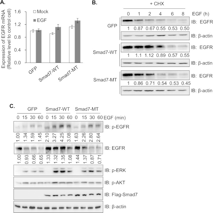
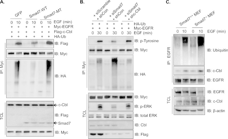
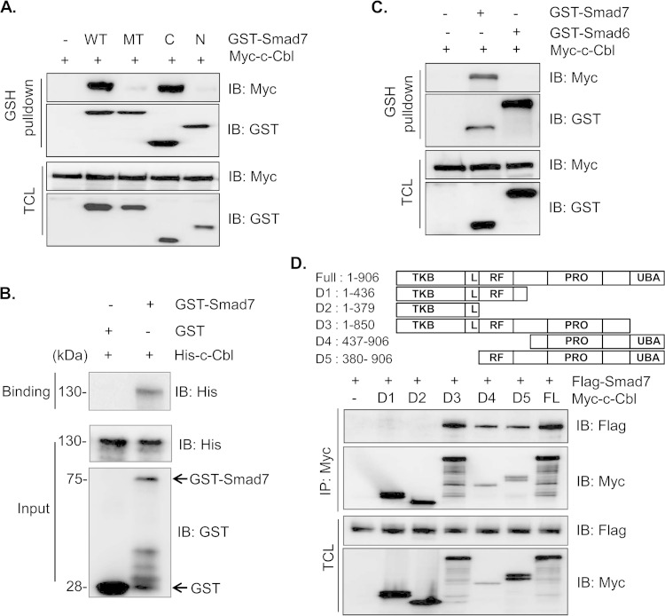
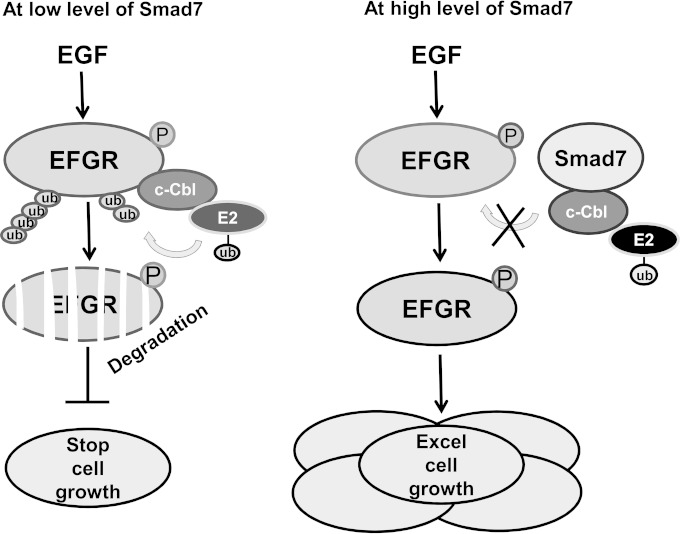
Epidermal growth factor (EGF) regulates various cellular events, including proliferation, differentiation, migration, and tumorigenesis. For the maintenance of homeostasis, EGF signaling should be tightly regulated to prevent the aberrant activation. Smad7 has been known as inhibitory Smad that blocks the signal transduction of transforming growth factor β. In the process of cell proliferation or transformation, Smad7 has been shown the opposite activities as a promoter or suppressor depending on cell types or microenvironments. We found that the overexpression of Smad7 in human HaCaT keratinocyte cells and mouse skin tissues elevated EGF receptor (EGFR) activity by impairing ligand-induced ubiquitination and degradation of activated receptor, which is induced by the E3 ubiquitin ligase c-Cbl. The C-terminal MH2 region but not MH1 region of Smad7 is critical for interaction with c-Cbl to inhibit the ubiquitination of EGFR. Interestingly, wild-type Smad7, but not Smad6 or mutant Smad7, destabilized the EGF-induced complex formation of c-Cbl and EGFR. These data suggest a novel role for Smad7 as a promoter for prolonging the EGFR signal in keratinocyte and skin tissue by reducing its ligand-induced ubiquitination and degradation.


Figures

FIG 1
FIG 1
Enhanced activity of EGF signaling by Smad7. (A) HaCaT control, Smad7 wild-type (WT), or Smad7 mutant (MT) cells were treated with various concentrations of EGF. After 3 days, cell numbers were measured as described in Materials and Methods. The results are representative of at least three independent experiments. (B) HaCaT control, Smad7-WT, or Smad7-MT cells were incubated with or without cetuximab (Cetu; 5 μg/ml) for 2 h after treatment with EGF (20 ng/ml) for 4 h. The mRNA levels of c-Myc and CCND1 were examined by qRT-PCR. The expression level of each mRNA was normalized based on cyclophilin mRNA expression. The results are representative of at least three independent experiments. *, P < 0.05 (Student t test). (C) HaCaT control, Smad7-WT, or Smad7-MT cells were plated and stimulated with EGF and cextuximab as described in Materials and Methods. After 2 weeks, the colonies that formed were counted and compared between samples. Representative phase-contrast images are shown. *, P < 0.05 (Student t test).
FIG 2
FIG 2
Increased stability of EGFR protein by Smad7. (A) To check the effect of Smad7 on the expression level of EGFR mRNA, HaCaT control, Smad7-WT, or Smad7-MT cells were treated with EGF (20 ng/ml) for 4 h. EGFR mRNA expression levels were detected by qRT-PCR. The mRNA expression levels of EGFR were normalized based on cyclophilin mRNA expression. The results are representative of at least three independent experiments. (B) To examine the protective activity of Smad7 on stability of the EGFR protein, control and Smad7-WT- or Smad7-MT-overexpressing HaCaT cells were starved for 24 h to allow maximum expression of EGFR. The cells were pretreated with cycloheximide (CHX) for 1 h after stimulation with EGF (20 ng/ml) for various times or left untreated. Immunoblot analyses were performed with the relevant antibodies. The relative band intensity of EGFR was measured with densitometry. (C) To see the effect of Smad7 on EGFR signaling, control and Smad7-WT- or Smad7-MT-overexpressing HaCaT cells were starved for 24 h and stimulated with EGF (20 ng/ml) for 15, 30, and 60 min or left untreated. Immunoblot analyses were performed with the relevant antibodies. The β-actin was detected as loading control. Representative gels of at least three different experiments are shown. The relative band intensity of phosphorylated or total EGFR was measured using densitometry.
FIG 3
FIG 3
Inhibitory activity of Smad7 on the ubiquitination of EGFR. (A) To check the differences in ubiquitination pattern of EGFR, HEK293T cells were transiently transfected with control vector, Flag-Smad7-WT, or Flag-Smad7-MT in combination with Myc-EGFR and HA-ubiquitin (Ub). At 36 h posttransfection, the cells were starved and stimulated with EGF (20 ng/ml) for 30, 60, and 90 min. Cell lysates were immunoprecipitated with antibodies specific for Myc (Myc-EGFR) and monitored by immunoblotting with anti-HA (ubiquitin) or anti-Myc antibodies. Total cell lysates (TCL) were immunoblotted with the relative antibodies. (B) To confirm the involvement of Smad7 on ubiquitination of EGFR, Smad7-overexpressing HEK293T cells were transfected with siRNA targeting Smad7 or scramble oligonucleotide. After 8 h, the cell lines were cotransfected with Myc-EGFR and HA-ubiquitin. The cells were then starved and stimulated with EGF (20 ng/ml) for 15 and 30 min. The cell lysates were immunoprecipitated with antibodies specific for Myc-EGFR and immunoblotted with anti-HA (ubiquitin) or antiphosphotyrosine (p-tyrosine) antibodies. The total cell lysates were immunoblotted with the relative antibodies. Representative gels from at least three different experiments are shown.
FIG 4
FIG 4
Blocking of EGFR/c-Cbl complex formation by Smad7. (A) To identify the molecular mechanism of EGFR stability, HEK293T cells were transiently cotransfected with control vector, Flag-Smad7-WT, or Flag-Smad7-MT and with Myc-EGFR, Flag-c-Cbl, or HA-ubiquitin. At 36 h posttransfection, the cells were starved and pretreated with MG132 for 4 h and stimulated with EGF (20 ng/ml) for 10 min. The cell lysates were immunoprecipitated with antibody specific for Myc-EGFR, followed by immunoblotting with anti-Flag (c-Cbl), anti-HA (ubiquitin) and anti-Myc antibodies. The total cell lysates were immunoblotted with anti-Flag and anti-Myc antibodies to validate the expression of each protein. (B) Stably Smad7-expressing HEK293T cells were infected with lentiviral shRNA against c-Cbl and transfected with siRNA targeting Smad7 or scramble oligonucleotide. At 8 h posttransfection, the cells were cotransfected with Myc-EGFR and HA-ubiquitin and stimulated with EGF (20 ng/ml) for 30 min. The cell lysates were immunoprecipitated with antibodies specific for Myc-EGFR, followed by immunoblotting with anti-HA (ubiquitin) or anti-p-tyrosine antibodies. The total cell lysates were immunoblotted with relative antibodies. (C) To confirm the effect of endogenous Smad7, Smad7+/+ and Smad7−/− MEFs were starved and incubated for 10 min with EGF. The total EGFR was immunoprecipitated with anti-EGFR antibody, and ubiquitinated protein was detected with antiubiquitin antibody. The total cell lysates were checked with the indicated antibodies to confirm the expression of each protein.
FIG 5
FIG 5
Binding domain mapping of Smad7 and c-Cbl. (A) To map the binding domain of Smad7, plasmids encoding GST-fused Smad7-WT, Smad7-MT, N-terminal (N), or C-terminal (C) fragments were transfected with Myc-tagged c-Cbl in to HEK293T cells. The cell lysates were immunoprecipitated with glutathione (GSH) beads, and interacted proteins were detected with anti-Myc and GST antibodies. (B) Recombinant GST or GST-Smad7 was incubated with equal amounts of His–c-Cbl fusion protein. After pulldown with GSH-beads, the bound proteins were immunoblotted with anti-His antibody. GST and GST-Smad7 were detected by immunoblotting with anti-GST antibody. (C) To confirm the specificity of Smad7 on interaction with c-Cbl, plasmids encoding GST-fused Smad7 or Smad6 were transfected with Myc-tagged c-Cbl into HEK293T cells. Cell lysates were immunoprecipitated with GSH beads, and interacted proteins were detected with anti-Myc and GST antibodies. (D) To map the binding site of c-Cbl, various deletion constructs were cotransfected into HEK293T cells with Smad7. Coimmunoprecipitation assay was performed with anti-Myc (c-Cbl) antibody, and immunoprecipitated proteins were detected with anti-Flag antibody. TKB, tyrosine kinase-binding domain; L, linker domain; RF, RING finger type E3 ligase domain; PRO, proline-rich domain; UBA, ubiquitin-associated domain.
FIG 6
FIG 6
In vivo validation of Smad7 activity on EGF signaling. (A) Groups of mice expressing Smad7-WT or MT on the skin were treated with TPA for the indicated times. The protein lysates were prepared from skin tissues taken from the backs of mice. Immunodetection of the total EGFR, the p-EGFR, the downstream effectors, and the p-AKT was performed using specific antibodies. β-Actin was detected as a loading control. (B) To check the effect of Smad7 on stability or ubiquitination of EGFR, skin lysates after TPA treatment were immunoprecipitated with antibodies specific for EGFR, followed by immunoblotting with antiubiquitin, antiphosphotyrosine, and anti-EGFR antibodies. The total cell lysates were immunoblotted with relative antibodies. (C) To confirm the in vivo interaction of Smad7 and c-Cbl, protein lysates after TPA treatment were immunoprecipitated with antibody specific for Smad7, followed by immunoblotting with anti-c-Cbl or anti-Smad7 antibodies. The total cell lysates were immunoblotted with the relative antibodies. Representative gels from at least three different experiments are shown.
FIG 7
FIG 7
Schematic diagram for the promoting activity of Smad7 on EGF signaling. In low levels or the absence of Smad7, c-Cbl upon EGF stimulation binds directly or indirectly to activated EGFR. The association of c-Cbl with EGFR results in activation of the E3 ligase activity of c-Cbl, which then targets EGFR for ubiquitination and degradation. In high levels or the presence of Smad7, c-Cbl proteins are sequestered far away from EGFR by the interaction between Smad7 and c-Cbl. The disruption of c-Cbl/EGFR association could enhance the activation of EGFR signaling, leading to the activation of its downstream signals, which contributes to cell proliferation and tumor formation.

References

    1. Derynck R, Akhurst RJ. 2007. Differentiation plasticity regulated by TGF-beta family proteins in development and disease. Nat Cell Biol 9:1000–1004. doi:10.1038/ncb434. - DOI - PubMed
    1. Massagué J. 2008. TGFβ in Cancer Cell 134:215–230. doi:10.1016/j.cell.2008.07.001. - DOI - PMC - PubMed
    1. Nakao A, Afrakhte M, Morén A, Nakayama T, Christian JL, Heuchel R, Itoh S, Kawabata M, Heldin NE, Heldin CH, ten Dijke P. 1997. Identification of Smad7, a TGF-β-inducible antagonist of TGF-β signalling. Nature 389:631–635. doi:10.1038/39369. - DOI - PubMed
    1. Ebisawa T, Fukuchi M, Murakami G, Chiba T, Tanaka K, Imamura T, Miyazono K. 2001. Smurf1 interacts with transforming growth factor-beta type I receptor through Smad7 and induces receptor degradation. J Biol Chem 276:12477–12480. doi:10.1074/jbc.C100008200. - DOI - PubMed
    1. Zhang S, Fei T, Zhang L, Zhang R, Chen F, Ning Y, Han Y, Feng X-H, Meng A, Chen Y-G. 2007. Smad7 antagonizes transforming growth factor beta signaling in the nucleus by interfering with functional Smad-DNA complex formation. Mol Cell Biol 27:4488–4499. doi:10.1128/MCB.01636-06. - DOI - PMC - PubMed

Publication types

MeSH terms

LinkOut - more resources