. 2015 Aug;35(16):2831-40.
doi: 10.1128/MCB.00263-15. Epub 2015 Jun 8.

A Region of Bdp1 Necessary for Transcription Initiation That Is Located within the RNA Polymerase III Active Site Cleft

Affiliations

A Region of Bdp1 Necessary for Transcription Initiation That Is Located within the RNA Polymerase III Active Site Cleft

Hui-Lan Hu et al. Mol Cell Biol. 2015 Aug.

Abstract

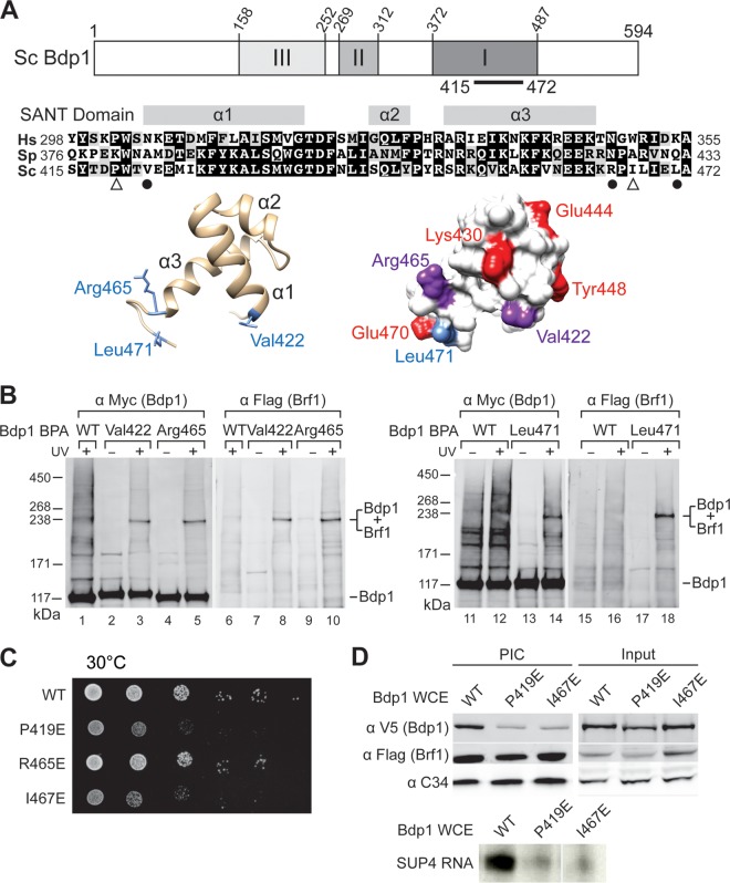
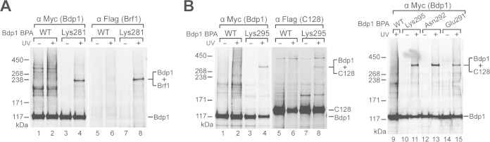
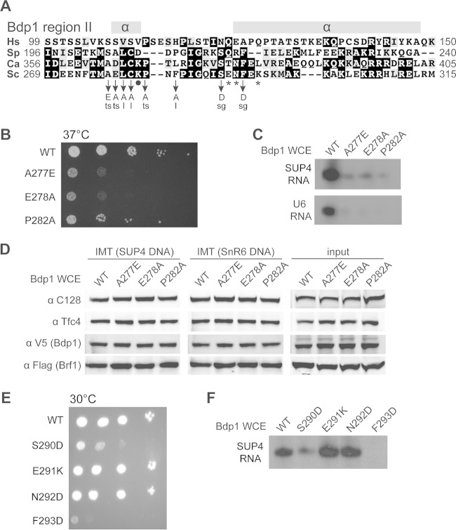
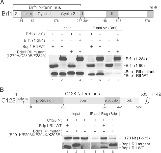
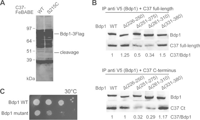
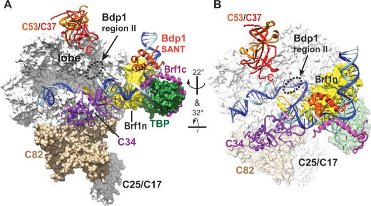
The RNA polymerase III (Pol III)-specific transcription factor Bdp1 is crucial to Pol III recruitment and promoter opening in transcription initiation, yet structural information is sparse. To examine its protein-binding targets within the preinitiation complex at the residue level, photoreactive amino acids were introduced into Saccharomyces cerevisiae Bdp1. Mutations within the highly conserved SANT domain cross-linked to the transcription factor IIB (TFIIB)-related transcription factor Brf1, consistent with the findings of previous studies. In addition, we identified an essential N-terminal region that cross-linked with the Pol III catalytic subunit C128 as well as Brf1. Closer examination revealed that this region interacted with the C128 N-terminal region, the N-terminal half of Brf1, and the C-terminal domain of the C37 subunit, together positioning this region within the active site cleft of the preinitiation complex. With our functional data, our analyses identified an essential region of Bdp1 that is positioned within the active site cleft of Pol III and necessary for transcription initiation.


Figures

FIG 1
FIG 1
The Bdp1 SANT domain cross-links to Brf1 and functions in PIC formation. (A) (Top) Essential regions I, II, and III in Saccharomyces cerevisiae (Sc) Bdp1 are shaded in the schematic. The black bar indicates the SANT domain. (Middle) Sequence alignment of the SANT domain from Bdp1 homologues. Hs, Homo sapiens (UniProt [http://www.uniprot.org/] accession number A6H8Y1; Sp, Schizosaccharomyces pombe, accession number O94481; Sc, Saccharomyces cerevisiae, UniProt accession number P46678. The three α helices in the SANT domain are indicated. Identical and similar residues are shaded in black and gray, respectively. Symbols below the sequence alignment: dots, residues with BPA cross-linking to Brf1; triangles, point mutations resulting in slow cell growth and defective transcription activity. (Bottom) Structural model of the SANT domain in a backbone trace (left) and the molecular surface (right). The model was generated by the Modeler program with the sequence with PDB accession number 2CU7 as the template. The blue side chains in the backbone trace model indicate residues involved in cross-linking with Brf1. In the molecular surface model, red residues are proposed to directly interact with Brf1, on the basis of NMR analysis. The residues interacting with Brf1, derived from both NMR analysis and BPA cross-linking, are in purple. Leu471 is proposed to interact only with Brf1 on the basis of BPA cross-linking. (B) Western analysis of photo-cross-linking. The BPA-substituted residues in the SANT domain are indicated above the lanes. Anti-Myc antibody revealed Myc epitope-tagged Bdp1 and fusion protein (lanes 1 to 5 and lanes 11 to 14). Anti-Flag antibody revealed the Flag epitope-tagged Brf1 in the fusion protein (lanes 6 to 10 and lanes 15 to 18). UV + and UV −, UV irradiation and no UV irradiation, respectively; WT, wild-type Bdp1 without BPA. (C) Cell growth analysis of Bdp1 SANT domain mutants by serial dilution spot assay. Representative slow cell growth at 30°C is shown. (D) Bdp1 SANT domain mutations affect PIC formation. (Top) Western blot analysis of proteins isolated from PIC formation. As indicated, Bdp1 mutant or WT whole-cell extracts were used in the IMT assay to isolate PICs on DNA containing the SUP4 gene. Individual proteins were probed with the antibodies indicated on the left. (Bottom) Autoradiogram of the synthesized SUP4 preliminary tRNA from PICs of the IMT assay.
FIG 2
FIG 2
Bdp1 region II cross-links to Brf1 and C128. (A) Bdp1-Brf1 photo-cross-linking. Bdp1 and the photo-cross-linked bands were detected by anti-Myc antibody (lanes 1 to 4). Anti-Flag antibody against tagged Brf1 confirmed its presence in the cross-linked polypeptide (lanes 5 to 8). (B) BPA-substituted residues Glu291, Asn292, and Lys295 in Bdp1 cross-linked with C128 (right), as confirmed by probing with anti-Flag antibody against Flag-tagged C128 in the fusion protein band (left).
FIG 3
FIG 3
Functional analysis of Bdp1 region II. (A) Bdp1 region II multiple-sequence alignment. As depicted in Fig. 1A, region II of Saccharomyces cerevisiae (Sc) Bdp1 ranges from amino acids 269 to 315. Hs, Homo sapiens; Sp, Schizosaccharomyces pombe; Ca, Candida albicans. The secondary structure of region II was predicted by use of the PSIPRED protein analysis workbench (http://bioinf.cs.ucl.ac.uk/psipred/). Symbols below the sequence alignment: asterisks, residues with BPA-cross-linking to C128; dot, the residue with BPA-cross-linking to Brf1. Point mutations and associated cell growth phenotypes are listed below. ts, temperature-sensitive (37°C) phenotype; sg, slow growth at all temperatures (16, 25, 30, and 37°C); l, amino acid substitution causing lethality. (B) Cell growth analysis of mutants with mutations in Bdp1 region II residues flanking the Brf1 BPA-cross-linking site. The three mutations caused a temperature-sensitive phenotype, as shown by serial dilution cell growth at 37°C. (C) In vitro transcription analysis for Bdp1 region II mutations flanking the residue cross-linking to Brf1. The autoradiograms show RNAs generated from the PICs of the IMT assay. As indicated, whole-cell extracts containing either WT or Bdp1 mutants were utilized in the in vitro transcription analysis. (D) PIC formation analysis of Bdp1 region II mutants. As indicated, Bdp1 mutant or WT whole-cell extracts were used in the IMT assay to isolate PICs on DNAs containing the SUP4 or SnR6 (U6) gene. Individual proteins of the isolated PICs were probed with antibodies, as indicated, in the Western blot analysis. (E) Cell growth analysis of mutations in Bdp1 region II residues involved in the C128 interaction. Representative slow cell growth at 30°C is shown. (F) In vitro transcription analysis for mutations in Bdp1 region II residues involved in the C128 interaction. The autoradiogram shows SUP4 tRNA from an in vitro transcription assay using WT or Bdp1 mutant whole-cell extracts.
FIG 4
FIG 4
Bdp1 region II interacts with the Brf1 N terminus and the C128 N terminus. (A) (Top) A schematic of Brf1 indicates the N-terminal zinc ribbon, linker, and cyclin fold repeat domains as well as the C-terminal conserved sequence blocks I, II, and III. (Bottom) Western blot analysis of coimmunoprecipitation with V5 epitope-tagged Brf1 N-terminal fragments from aa 1 to 90 and aa 1 to 284 and a Bdp1 region II peptide fragment containing aa 253 to 325. Anti-V5 antibody-agarose beads were used to precipitate the V5-tagged Brf1 fragments. As indicated, either the WT Bdp1 region II peptide or the mutant peptide with triple point mutations was applied in the coimmunoprecipitation assay. (B) (Top) A schematic of the C128 N-terminal region includes the external 1, protrusion, lobe, and fork domains. (Bottom) Western blot analysis of coimmunoprecipitation with Bdp1 region II and C128 N-terminal (Nt) peptide fragments. Anti-Flag antibody-agarose beads were applied to precipitate the Flag-tagged Bdp1 peptide fragment. As indicated, either the WT Bdp1 region II fragment or the Bdp1 region II fragment with a quadruple point mutation was used. Coimmunoprecipitated polypeptides were probed with anti-Flag and anti-His antibodies, revealing the Bdp1 region and the C128 N terminus, respectively.
FIG 5
FIG 5
The C37 C terminus interacts with Bdp1 region II. (A) Western blot analysis of hydroxyl radical cleavage of Bdp1 by FeBABE-conjugated C37. As indicated, either the wild type or the C37 mutant with a single cysteine mutation at Ser215 was used in the FeBABE conjugation and subsequent hydroxyl radical protein cleavage assay. C-terminally Flag-tagged full-length Bdp1 and the cleaved peptide fragments were visualized by probing with an anti-Flag antibody. The corresponding cleavage site for the Bdp1 peptide fragment is aa 264. (B) Western blot analysis of coimmunoprecipitation of Bdp1 and C37. Anti-V5 antibody beads were used to precipitate the C-terminally V5 epitope-tagged Bdp1 and internal deletion mutants. Either the C37 full-length protein (top) or the C37 C-terminal (Ct) peptide fragment from aa 181 to 281 (bottom) was used in the coimmunoprecipitation assay. Bdp1 and C37 were probed with anti-V5 and anti-His antibodies, respectively. The relative intensity of C37 immunostaining is normalized to the intensity of Bdp1 immunostaining and is listed below each lane. (C) The cell growth phenotype was analyzed by a serial dilution spot assay. As indicated, Bdp1 quadruple point mutant R260E/D261K/E263K/K266E and WT strains were grown at 30°C.
FIG 6
FIG 6
Positions of Bdp1 region II and the SANT domain in the Pol III open promoter complex model. (A) The model of the Pol III-Brf1-TBP-DNA open promoter complex was initially assembled with the structures of the Pol II-TFIIB-TBP complex, the Brf1-TBP-DNA complex, and Pol I (31, 43–46). The C82 and C34 structures were generated on the basis of homology modeling using structures of human homologues as the templates. The C53/C37 dimerization module was generated using the human TFIIF (Rap74/Rap30) dimer structure as the homology modeling template. C82, C34, and the C53/C37 dimerization module were added to Pol III on the basis of biochemical cross-linking and binding data, as described previously (31). Template and nontemplate DNA strands are in blue and cyan, respectively. The Pol III 12-subunit core structure (white), C82 (tan), the Brf1 cyclin fold repeat domain (yellow), and TBP (dark green) are shown as a molecular surface representation. The magenta sphere in the Pol III core structure represents the active site magnesium ion. C34 (purple), C53 (orange), C37 (red), the Bdp1 SANT domain (orange-red), and Brf1 C-terminal block II (magenta) are shown as backbone trace representations. TFIIB N-terminal zinc ribbon, reader, and linker domains (yellow) are placed in the Pol III active site cleft to indicate likely positions for Brf1 N-terminal zinc ribbon and linker domains. (B) Different orientation of the open promoter complex. Pol III core, C82, and TBP are in semitransparent surface representations.

References

    1. Dieci G, Fiorino G, Castelnuovo M, Teichmann M, Pagano A. 2007. The expanding RNA polymerase III transcriptome. Trends Genet 23:614–622. doi:10.1016/j.tig.2007.09.001. - DOI - PubMed
    1. Vannini A, Cramer P. 2012. Conservation between the RNA polymerase I, II, and III transcription initiation machineries. Mol Cell 45:439–446. doi:10.1016/j.molcel.2012.01.023. - DOI - PubMed
    1. Geiduschek EP, Kassavetis GA. 2001. The RNA polymerase III transcription apparatus. J Mol Biol 310:1–26. doi:10.1006/jmbi.2001.4732. - DOI - PubMed
    1. Schramm L, Hernandez N. 2002. Recruitment of RNA polymerase III to its target promoters. Genes Dev 16:2593–2620. doi:10.1101/gad.1018902. - DOI - PubMed
    1. Kassavetis GA, Bartholomew B, Blanco JA, Johnson TE, Geiduschek EP. 1991. Two essential components of the Saccharomyces cerevisiae transcription factor TFIIIB: transcription and DNA-binding properties. Proc Natl Acad Sci U S A 88:7308–7312. doi:10.1073/pnas.88.16.7308. - DOI - PMC - PubMed

Publication types

MeSH terms

Associated data

LinkOut - more resources