. 2015 Aug;35(16):2851-63.
doi: 10.1128/MCB.01064-14. Epub 2015 Jun 8.

TRIM28 Is an E3 Ligase for ARF-Mediated NPM1/B23 SUMOylation That Represses Centrosome Amplification

Affiliations

TRIM28 Is an E3 Ligase for ARF-Mediated NPM1/B23 SUMOylation That Represses Centrosome Amplification

Shu Hui Neo et al. Mol Cell Biol. 2015 Aug.

Abstract

The tumor suppressor ARF enhances the SUMOylation of target proteins; however, the physiological function of ARF-mediated SUMOylation has been unclear due to the lack of a known, associated E3 SUMO ligase. Here we uncover TRIM28/KAP1 as a novel ARF-binding protein and SUMO E3 ligase for NPM1/B23. ARF and TRIM28 cooperate to SUMOylate NPM1, a nucleolar protein that regulates centrosome duplication and genomic stability. ARF-mediated SUMOylation of NPM1 was attenuated by TRIM28 depletion and enhanced by TRIM28 overexpression. Coexpression of ARF and TRIM28 promoted NPM1 centrosomal localization by enhancing its SUMOylation and suppressed centrosome amplification; these functions required the E3 ligase activity of TRIM28. Conversely, depletion of ARF or TRIM28 increased centrosome amplification. ARF also counteracted oncogenic Ras-induced centrosome amplification. Centrosome amplification is often induced by oncogenic insults, leading to genomic instability. However, the mechanisms employed by tumor suppressors to protect the genome are poorly understood. Our findings suggest a novel role for ARF in maintaining genome integrity by facilitating TRIM28-mediated SUMOylation of NPM1, thus preventing centrosome amplification.


Figures

FIG 1
FIG 1
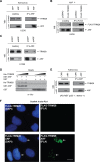
TRIM28 is a novel binding partner of ARF. (A and B) Ectopically expressed ARF binds TRIM28. U2OS cells were infected with adenoviruses encoding the indicated proteins (A) or transfected with the indicated plasmids (B). Immunoprecipitation (IP) was performed with the indicated antibodies and analyzed by Western blotting (WB). (C) ARF binds endogenous TRIM28. Lysates of H1299 cells expressing retrovirus-encoded control GFP short hairpin RNA (shRNA) or ARF shRNA were immunoprecipitated with anti-ARF antibody. (D) Direct interaction between ARF and TRIM28. Purified bacterial recombinant His6-TRIM28 was mixed with GST-ARF, followed by pulldown with glutathione beads. The resulting complexes were analyzed by Western blotting. (E) ARF binds TRIM28 independently of both p53 and MDM2. Cell lysates from Ad-ARF-infected p53−/−; Mdm2−/− MEFs were immunoprecipitated with anti-ARF antibody. (F) ARF binds TRIM28 in the nucleus. U2OS cells were transfected with the indicated plasmids and analyzed by Duolink PLA. Cells with positive PLA signals (green [indicated by arrows], right panels) indicates ARF-TRIM28 binding. Left panels show DAPI-stained nuclei. Scale bar, 3 μm.
FIG 2
FIG 2
Domain analysis of the ARF-TRIM28 interaction. Mapping of the TRIM28-binding domain of ARF (A) and ARF-binding domain of TRIM28 (B). Lysates of U2OS cells transfected with indicated plasmids were immunoprecipitated with anti-Myc antibody and analyzed by Western blotting (WB) with the indicated antibodies.
FIG 3
FIG 3
TRIM28 is an E3 ligase for ARF-mediated SUMOylation of NPM1. (A) In vitro SUMOylation assay. NPM1 is SUMOylated by TRIM28, and ARF enhances this reaction. The relative band intensities of the SUMOylated NPM1 normalized to non-SUMOylated NPM1 (SUMO-NPM1/NPM1) are shown. The value for lane 3 is set at 1.0. (B) Depletion of ARF reduces SUMOylation of NPM1. Adenovirus (Ad)-encoded TRIM28-infected H1299 cells expressing either GFP or ARF shRNA were transfected with the indicated plasmids. SUMOylated proteins were recovered with Ni2+-NTA beads and analyzed by Western blotting. The ratio of SUMO-NPM1 to NPM1 is shown as described for panel A. The value of SUMO-NPM1/NPM1 for lane 3 is set at 1.0. (C) Loss of ARF reduces SUMOylation of NPM1. The experiment was performed as described for panel B using early-passage p53−/− and p53−/−; ARF−/− MEFs. The value of SUMO-NPM1/NPM1 for lane 2 is set at 1.0. (D) Depletion of ARF reduces SUMOylation of NPM1. The experiment was performed as described for panel B using Saos-2 cells expressing either GFP or ARF shRNA. The value of SUMO-NPM1/NPM1 for lane 2 is set at 1.0. (E) Overexpression of both TRIM28 and ARF dramatically increases SUMOylation of NPM1. Adenovirus (Ad)-encoded TRIM28-infected H1299 cells were transfected with the indicated plasmids and analyzed as described for panel B. The value of SUMO-NPM1/NPM1 for lane 2 is set at 1.0. (F) Depletion of TRIM28 reduces SUMOylation of NPM1. The experiment was performed as described for panel B using H1299 cells, with the exception that TRIM28 expression was knocked down by siRNA. The value of SUMO-NPM1/NPM1 for lane 1 is set at 1.0. (G) NPM1 SUMOylation induced by overexpression of ARF is reduced by knockdown of TRIM28. The experiment was performed as described for panel B using U2OS cells, with the exception that TRIM28 expression was knocked down by siRNA. The value of SUMO-NPM1/NPM1 for lane 1 is set at 1.0. (H) The reduction of NPM1 SUMOylation by knockdown of ARF is attenuated by overexpression of TRIM28. The experiment was performed as described for panel B using H1299 cells expressing either GFP or ARF shRNA. The value of SUMO-NPM1/NPM1 for lane 1 is set at 1.0.
FIG 4
FIG 4
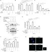
ARF represses centrosome amplification. (A) Loss of ARF results in enhanced centrosome amplification. Representative images (left) of wild-type and early-passage ARF−/− MEFs immunostained with anti-γ-tubulin antibody (green) and stained with DAPI (blue). Centrosomes (γ-tubulin-positive foci) were counted and plotted (right). Scale bar, 2 μm. (B) Depletion of ARF in human cancer cells increases centrosome amplification. Centrosomes in Saos-2 cells expressing either shGFP or shARF were quantified as described for panel A. Scale bar, 2 μm. (C) Overexpression of ARF but not ARF deletion mutant ARF(65-132), which lacks the TRIM28-binding region, decreases centrosome amplification. ARF−/− MEFs were transfected with the indicated plasmids, and centrosomes (γ-tubulin-positive foci) were counted as described for panel A. (D) Overexpression of ARF but not ARF deletion mutant ARF(65-132) decreases centrosome amplification. U2OS cells were transfected with the indicated plasmids, and centrosomes (γ-tubulin-positive foci) were counted as described for panel A. (E and F) Oncogenic Ras-induced centrosome amplification was attenuated by ARF. Early-passage p53−/− and p53−/−; ARF−/− MEFs were infected with empty or (V12) Ras-expressing retroviruses. Cells were selected using puromycin, and cell lysates were harvested and analyzed by Western blotting (E). Histone H3 serves as the loading control. Centrosome numbers were counted as described for panel A (F). Data in panels A to D and F were determined by counting ≥200 cells. Data are represented as means ± standard deviations from three independent experiments. Statistical significance: *, P ≤ 0.05; **, P ≤ 0.01; ***, P ≤ 0.001; ns, not significant.
FIG 5
FIG 5
TRIM28 contributes to ARF-mediated repression of centrosome amplification. (A) TRIM28 and ARF contribute to inhibition of centrosome amplification. Saos-2 cells expressing shGFP or shARF were transfected with TRIM28 siRNA and stained with anti-γ-tubulin. Centrosome numbers were counted as described for Fig. 4. (B) Reduction of centrosome amplification by ARF overexpression is attenuated by knockdown of TRIM28. Saos-2 cells expressing shARF were transfected with the indicated plasmids and siRNA and stained with anti-γ-tubulin. Centrosome numbers were counted as described for Fig. 4. (C) TRIM28 contributes to the reduction of centrosome amplification even in shARF-expressing cells. Saos-2 cells expressing shGFP or shARF were transfected with FLAG-TRIM28 expressing plasmid and stained with anti-γ-tubulin. Centrosome numbers were counted as described for Fig. 4. (D) Wild-type TRIM28, but not a catalytic dead mutant TRIM28 C651F, enhances SUMOylation of NPM1 in the presence of ARF. NIH 3T3 cells were transfected with the indicated plasmids, recovered with Ni2+-NTA beads, and analyzed by Western blotting. The relative band intensities of the SUMOylated NPM1 normalized to non-SUMOylated NPM1 (SUMO-NPM1/NPM1) are shown. The value for lane 2 is set at 1.0. (E) ARF and TRIM28 reduce centrosome amplification dependent on E3 SUMO ligase activity of TRIM28. NIH 3T3 cells were cotransfected with the indicated plasmids and stained with anti-γ-tubulin. Centrosome numbers were determined as described for Fig. 4. (F and G) ARF and TRIM28 enhance centrosomal translocation of NPM1 dependent on E3 SUMO ligase activity of TRIM28. NIH 3T3 cells were transfected with the indicated plasmids, and colocalization of NPM1 and γ-tubulin was assessed by immunostaining. The percentage of NPM1 colocalized with γ-tubulin in transfected cells was plotted (F); representative images of colocalization between NPM1 and γ-tubulin are shown (G). Arrows indicate the positions of centrosomes. Scale bar, 2 μm. The field enclosed by white dashed lines is shown at a higher magnification to the right of merged images. More than 200 cells in panels A, B, and C and more than 300 cells in panels E and F were counted. Data are represented as means ± standard deviations from three independent experiments. Statistical significance: *, P ≤ 0.05; **, P ≤ 0.01; ***, P ≤ 0.001; ns, not significant.
FIG 6
FIG 6
Model of ARF-mediated NPM1 SUMOylation in preventing centrosome amplification. ARF prevents centrosome amplification induced by oncogenic insults by enhancing SUMOylation of NPM1 via TRIM28. The ARF-TRIM28 interaction promotes NPM1 SUMOylation, enhancing its translocation to centrosomes, thereby preventing centrosome amplification induced by oncogenic insults.

References

    1. Palmero I, Pantoja C, Serrano M. 1998. p19ARF links the tumour suppressor p53 to Ras. Nature 395:125–126. doi:10.1038/25870. - DOI - PubMed
    1. Sherr CJ. 2006. Divorcing ARF and p53: an unsettled case. Nat Rev Cancer 6:663–673. doi:10.1038/nrc1954. - DOI - PubMed
    1. Weber JD, Jeffers JR, Rehg JE, Randle DH, Lozano G, Roussel MF, Sherr CJ, Zambetti GP. 2000. p53-independent functions of the p19ARF tumor suppressor. Genes Dev 14:2358–2365. doi:10.1101/gad.827300. - DOI - PMC - PubMed
    1. Xirodimas DP, Chisholm J, Desterro JMS, Lane DP, Hay RT. 2002. P14ARF promotes accumulation of SUMO-1 conjugated (H)Mdm2. FEBS Lett 528:207–211. doi:10.1016/S0014-5793(02)03310-0. - DOI - PubMed
    1. Tago K, Chiocca S, Sherr CJ. 2005. Sumoylation induced by the Arf tumor suppressor: a p53-independent function. Proc Natl Acad Sci U S A 102:7689–7694. doi:10.1073/pnas.0502978102. - DOI - PMC - PubMed

Publication types

MeSH terms

LinkOut - more resources