Review
. 2015 Jul;11(7):394-410.
doi: 10.1038/nrneph.2015.82. Epub 2015 Jun 9.

Anaemia in kidney disease: harnessing hypoxia responses for therapy

Affiliations
Review

Anaemia in kidney disease: harnessing hypoxia responses for therapy

Mark J Koury et al. Nat Rev Nephrol. 2015 Jul.

Abstract

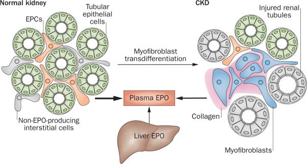
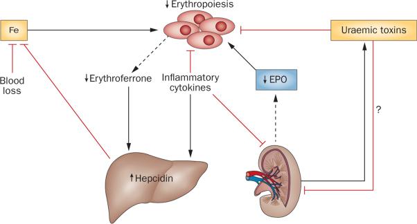
Improved understanding of the oxygen-dependent regulation of erythropoiesis has provided new insights into the pathogenesis of anaemia associated with renal failure and has led to the development of novel therapeutic agents for its treatment. Hypoxia-inducible factor (HIF)-2 is a key regulator of erythropoiesis and iron metabolism. HIF-2 is activated by hypoxic conditions and controls the production of erythropoietin by renal peritubular interstitial fibroblast-like cells and hepatocytes. In anaemia associated with renal disease, erythropoiesis is suppressed due to inadequate erythropoietin production in the kidney, inflammation and iron deficiency; however, pharmacologic agents that activate the HIF axis could provide a physiologic approach to the treatment of renal anaemia by mimicking hypoxia responses that coordinate erythropoiesis with iron metabolism. This Review discusses the functional inter-relationships between erythropoietin, iron and inflammatory mediators under physiologic conditions and in relation to the pathogenesis of renal anaemia, as well as recent insights into the molecular and cellular basis of erythropoietin production in the kidney. It furthermore provides a detailed overview of current clinical experience with pharmacologic activators of HIF signalling as a novel comprehensive and physiologic approach to the treatment of anaemia.


Figures

Figure 1
Figure 1
Overview of erythropoiesis. a | Progressive stages of erythroid differentiation showing the relative sizes and presumed or known morphologic appearances of haematopoietic cells at various stages. The transcription factors PU.1 and GATA1 are important in determining whether HSCs will progress towards an erythroid or a non-erythroid fate, whereas KLF1 is important in determining whether MEPs will progress towards an erythroid or a megakaryocytic fate. PU.1 expression continues until the EPO-dependent stages, whereas GATA1 and KLF1 have important roles in differentiation throughout haemoglobin synthesis. Stages of haemoglobin synthesis show relative accumulations of haemoglobin as increasing intensity of red in the cytoplasm. The periods of EPO dependence ending at the early Baso EB stage and haemoglobin synthesis beginning in the late Baso EB stage do not overlap. b | An erythroblastic island. Erythroid cells from the CFU-E through to the RET stages are attached to a central macrophage. RETs detach from the macrophage before leaving the marrow and entering the blood. Two extruded nuclei created when RETs are formed from Ortho EBs are shown inside the central macrophage where they have been phagocytosed and will be degraded. Abbreviations: Baso, basophilic; BFU-E, burst-forming unit-erythroid; CFU-E, colony-forming unit-erythroid; EB, erythroblast; EPO, erythropoietin; EPOR, EPO receptor; HSC, pluripotent haematopoietic stem cell; MEP, bipotent megakaryocytic–erythroid progenitor; Ortho, orthochromatic; Poly, polychromatophilic; RBC, erythrocyte; RET, reticulocyte.
Figure 2
Figure 2
Regulation of HIF degradation by PHDs and hypoxic induction of erythropoietin. HIF controls a wide spectrum of tissue-specific and systemic hypoxia responses, with HIF-2 being the principal regulator of EPO transcription in vivo. The HIF oxygen-sensing machinery targets HIF-1α, HIF-2α and HIF-3α for proteasomal degradation by the VHL–E3-ubiquitin ligase complex. In the presence of oxygen HIF-α is hydroxylated at specific proline residues (Pro-OH) by PHD enzymes. Hydroxylation of HIF-α permits binding to the β-domain of VHL, which functions as the substrate recognition component of the VHL–E3-ubiquitin ligase complex. Under hypoxic conditions, HIF-α degradation is inhibited and HIF-α translocates to the nucleus, where it forms a heterodimer with ARNT and binds hypoxia regulatory elements in EPO, which contain the HIF consensus binding site 5'-RCGTG-3'. The hypoxic induction of EPO in the liver is mediated by the liver-inducibility element in the 3'-end of the EPO gene, whereas the hypoxic induction of renal EPO requires the kidney inducibility element located upstream of the EPO transcription start site. The putative DNA sequence of the kidney inducibility element is shown in lower case letters. HNF-4 is an important coregulator of EPO. Boxes depict EPO exons. EPO coding sequences and non-translated sequences are depicted in red and blue, respectively. The distance from the EPO transcription start site is indicated in kilobases. Abbreviations: 2OG, 2-oxoglutarate; ARNT, aryl hydrocarbon receptor nuclear translocator; EPO, erythropoietin gene; Fe2+, ferrous iron; HIF, hypoxia-inducible factor; HNF-4, hepatocyte nuclear factor 4; PHD, prolyl-4-hydroxylase domain.
Figure 3
Figure 3
HIF coordinates erythropoietin production with iron metabolism. HIF-2 stimulates renal and hepatic erythropoietin synthesis, which raises serum erythropoietin levels, stimulating erythropoiesis in the bone marrow. In the duodenum, DCYTB reduces Fe3+ to Fe2+, which then enters enterocytes via DMT1. DCYTB and DMT1 are both regulated by HIF-2. Iron is then released into the circulation via FPN, which is also HIF-2-inducible. In the circulation iron is transported in a complex with TF to the liver, bone marrow and other organs; cells of the reticuloendothelial system acquire iron through the phagocytosis of senescent red cells. TF is HIF-regulated, and hypoxia and/or pharmacologic PHD inhibition raises TF serum levels. Increased erythropoietic activity in the bone marrow produces GDF15 and erythroferrone, which suppress hepcidin in hepatocytes. Hepcidin suppression increases FPN expression on enterocytes, hepatocytes and macrophages, resulting in increased iron absorption and mobilization from internal stores. Inflammation stimulates hepcidin production in the liver and leads to reduced FPN expression and hypoferraemia. In addition to regulating hepcidin indirectly by stimulating erythropoiesis, in vitro studies suggest that HIF might also modulate hepcidin expression through the regulation of furin and transmembrane protease serine 6. Abbreviations: DCYTB, duodenal cytochrome b reductase 1; DMT1, divalent metal transporter-1; EPO, plasma erythropoietin; Fe2+, ferrous iron; Fe3+, ferric iron; FPN, ferroportin; GDF15, growth differentiation factor 15; HIF, hypoxia-inducible factor; TF, transferrin.
Figure 4
Figure 4
The number of EPCs regulates renal erythropoietin output. a | Under physiologic, non-stimulated conditions a small number of renal EPCs are responsible for renal erythropoietin output. The size of the EPC pool is regulated in an oxygen-dependent manner and increases under hypoxic conditions. The expansion of the EPC pool requires HIF-2 signalling, which is activated by hypoxia, pharmacologic PHD inhibition, or as a consequence of mutations in the oxygen-sensing pathway. b | Renal EPCs are derived from FOXD1-expressing stromal cells, and include interstitial fibroblast-like cells, pericytes and renin-producing cells. Renin-producing cells can be induced to synthesize erythropoietin under conditions of Vhl gene inactivation; their role in hypoxia-induced renal erythropoietin production is unclear. Abbreviations: EPC, erythropoietin-producing cell; EPO, erythropoietin; HIF-2, hypoxia-inducible factor-2; VSMC, vascular smooth muscle cell.
Figure 5
Figure 5
Cellular basis of erythropoietin deficiency in renal failure. In the normal kidney, EPCs are recruited from peritubular interstitial fibroblast-like cells and pericytes. Tubular epithelial cells do not produce EPO. Under conditions of injury, EPCs or interstitial cells with EPC potential transdifferentiate into myofibroblasts, which synthesize collagen and lose their ability to produce EPO. In CKD, EPC recruitment is impaired, resulting in reduced renal EPO output and the development of anaemia. Under conditions of severe hypoxia or in patients with advanced CKD, the liver contributes to plasma EPO levels. Abbreviations: CKD, chronic kidney disease; EPC, erythropoietin-producing cell; EPO, erythropoietin.
Figure 6
Figure 6
Mechanisms of renal anaemia. In renal anaemia, the ability of the kidney to produce EPO is impaired. Inflammatory cytokines suppress erythropoiesis in the bone marrow, EPO production in the kidney, and stimulate hepcidin production in the liver, which negatively affects iron absorption and mobilization. Hepcidin is also maintained at higher levels by decreased erythroferrone production, which is secondary to a reduction in erythroblast numbers due to EPO deficiency. In patients with advanced CKD, the liver contributes significantly to plasma EPO levels. The contribution of uraemic toxins to the pathogenesis of renal anaemia is only poorly understood. Uraemic toxins have been shown to suppress erythroid colony formation in vitro as well as EPO transcription in hepatoma cells, the latter indicating possible suppressive effects on hepatic and renal EPO production in vivo., Abbreviations: CKD, chronic kidney disease; EPO, erythropoietin; Fe, iron.

References

    1. Mancini E, et al. FOG-1 and GATA-1 act sequentially to specify definitive megakaryocytic and erythroid progenitors. EMBO J. 2012;31:351–365. - PMC - PubMed
    1. Siatecka M, Bieker JJ. The multifunctional role of EKLF/KLF1 during erythropoiesis. Blood. 2011;118:2044–2054. - PMC - PubMed
    1. Gregory CJ, Eaves AC. Human marrow cells capable of erythropoietic differentiation in vitro: definition of three erythroid colony responses. Blood. 1977;49:855–864. - PubMed
    1. Chasis JA, Mohandas N. Erythroblastic islands: niches for erythropoiesis. Blood. 2008;112:470–478. - PMC - PubMed
    1. Nagata S. Autoimmune diseases caused by defects in clearing dead cells and nuclei expelled from erythroid precursors. Immunol. Rev. 2007;220:237–250. - PubMed

Publication types

MeSH terms

Substances