

Unusual apocrine carcinoma with neuroendocrine differentiation: a cutaneous neoplasm may be analogous to neuroendocrine carcinoma with apocrine differentiation of breast
- PMID: 26055980
- PMCID: PMC4460766
- DOI: 10.1186/s13000-015-0302-4
Unusual apocrine carcinoma with neuroendocrine differentiation: a cutaneous neoplasm may be analogous to neuroendocrine carcinoma with apocrine differentiation of breast
Abstract
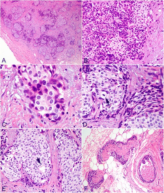
Cutaneous apocrine carcinoma (AC) is a rare adnexal neoplasm that histologically can mimic breast carcinoma metastatic to the skin or apocrine carcinoma arising in ectopic breast tissue. As extremely rare condition, neuroendocrine differentiation may be observed in AC although its etiology and pathogenesis is still unclear. We report here a case of unusual AC with neuroendocrine differentiation in right labium majus pudenda. A 43-year-old woman presented with a 6-month history of an asymptomatic pea-sized brownish nodule in right labium majus pudenda without enlargement of inguinal lymph nodes and bilateral breast nodules. The mass was totally resected. Microscopically, the tumor was solitary and located in the deep dermis without epidermal connection. Tumor cells were arranged in a micronodular or formed massive solid nests separated by densely fibroblastic stroma. Scattered glandular or rosette-like structures were identified within the tumor nodules. Immunohistochemically, the tumor cells were diffusely positive to CK7, CEA, GCDFP-15, synaptophysin, estrogen and progesterone receptors. Part of tumor cells expressed androgen receptor, but they were negative to CK20, CK5/6, p63 and S-100. Because of its rarity and histogenesis complexity, there exist diagnostic challenges for pathologists to differentiate cutaneous AC with neuroendocrine differentiation from other carcinomas with apocrine or neuroendocrine features. Our case demonstrates that the tumor shares some features with mammary carcinoma and might originate from mammary-like sweat gland in anogenital region. The results suggest that, for the first time, primary cutaneous AC with neuroendocrine differentiation may be analogous to the mammary neuroendocrine carcinoma with apocrine differentiation in histological feature and biological behavior. Virtual Slides: The virtual slide(s) for this article can be found here: http://www.diagnosticpathology.diagnomx.eu/vs/7732276716685708.
Figures

Comment in
-
Apocrine carcinoma with neuroendocrine differentiation.J Cutan Pathol. 2017 Sep;44(9):810-812. doi: 10.1111/cup.12977. Epub 2017 Jul 3. J Cutan Pathol. 2017. PMID: 28671291 No abstract available.
References
-
- Requena L, Kutzner H, Hurt MA, Santa Cruz DJ, Mehregan AH. Malignant tumors with apocrine and eccrine differentiation. In: LeBoit PE, Burg G, Weedon D, Sarasin A, editors. World Health Organization Classification of Tumours of Skin. Lyon: IARC press; 2006. pp. 125–38.
-
- Hayes MM, Matisic JP, Weir L. Apocrine carcinoma of the lip: a case report including immunohistochemical and ultrastructural study, discussion of differential diagnosis, and review of the literature. Oral Surg Oral Med Oral Pathol Oral Radiol Endod. 1996;82:193–9. doi: 10.1016/S1079-2104(96)80224-1. - DOI - PubMed
Publication types
MeSH terms
Substances
LinkOut - more resources
Full Text Sources
Other Literature Sources
Medical
Research Materials
